. 2013 Jan 15;110(3):E231-40.
doi: 10.1073/pnas.1206322110. Epub 2012 Dec 27.

Natural killer cells promote immune tolerance by regulating inflammatory TH17 cells at the human maternal-fetal interface

Affiliations

Natural killer cells promote immune tolerance by regulating inflammatory TH17 cells at the human maternal-fetal interface

Binqing Fu et al. Proc Natl Acad Sci U S A. .

Abstract

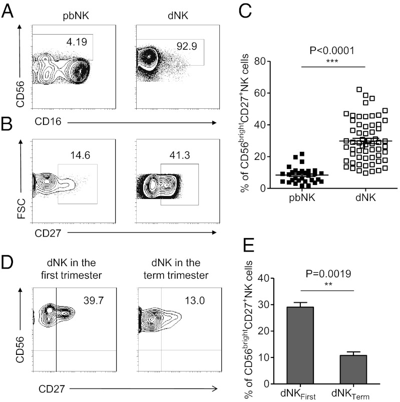
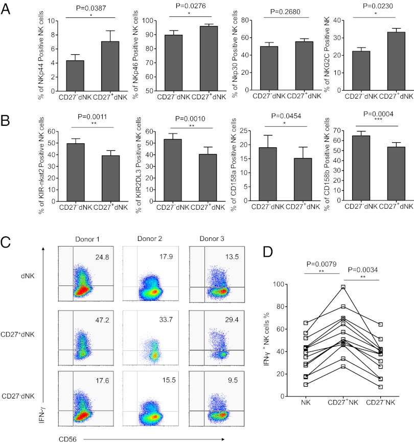
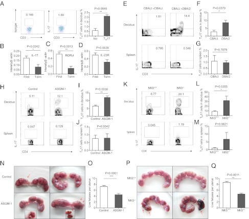
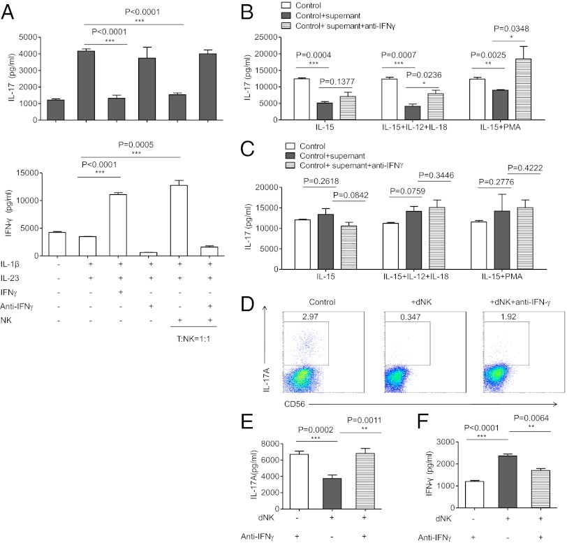
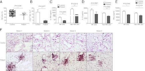
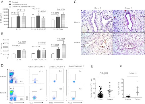
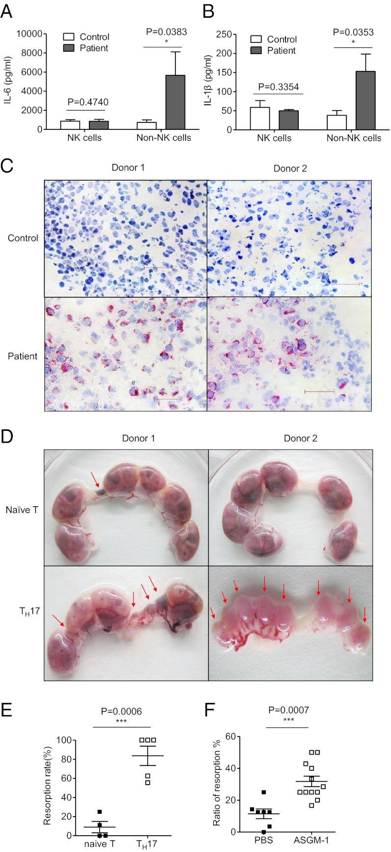
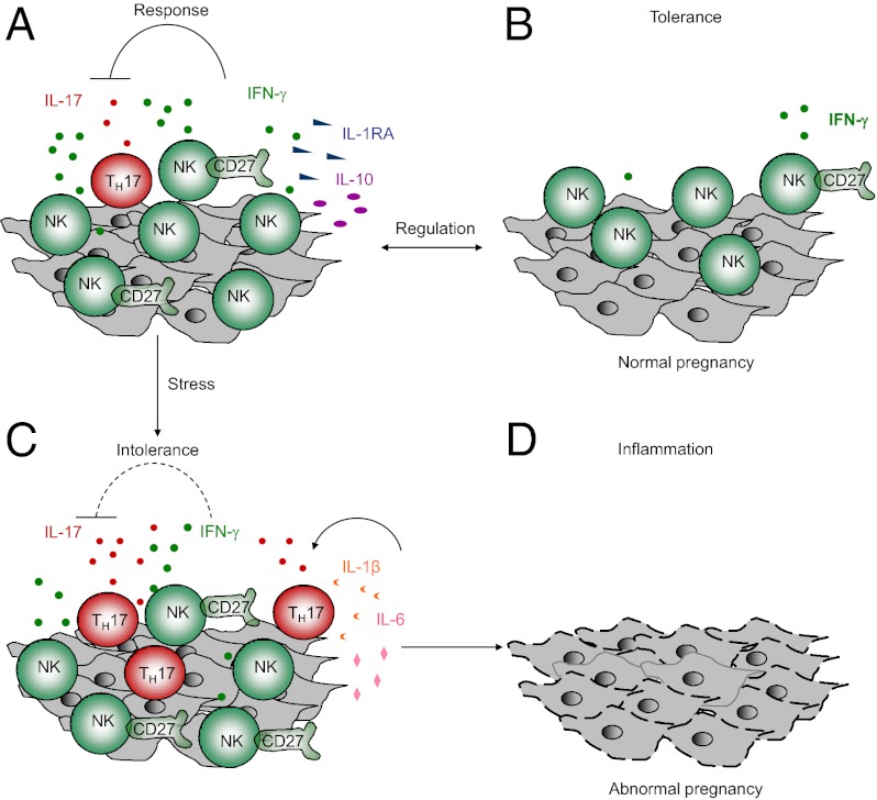
Natural killer (NK) cells accumulate at the maternal-fetal interface in large numbers, but their exact roles in successful pregnancy remain poorly defined. Here, we provide evidence that T(H)17 cells and local inflammation can occur at the maternal-fetal interface during natural allogenic pregnancies. We found that decidual NK cells promote immune tolerance and successful pregnancy by dampening inflammatory T(H)17 cells via IFN-γ secreted by the CD56(bright)CD27(+) NK subset. This NK-cell-mediated regulatory response is lost in patients who experience recurrent spontaneous abortions, which results in a prominent T(H)17 response and extensive local inflammation. This local inflammatory response further affects the regulatory function of NK cells, leading to the eventual loss of maternal-fetal tolerance. Thus, our data identify NK cells as key regulatory cells at the maternal-fetal interface by suppressing T(H)17-mediated local inflammation.


Conflict of interest statement

The authors declare no conflict of interest.

Figures

Fig. 1.
Fig. 1.
CD56brightCD27+ NK cells present in large numbers in human deciduas of the first trimester. (A) Representative density plots showing an analysis of CD56bright NK cells in gated CD56+CD3 NK cells isolated from peripheral blood (pbNK) and decidua in the first trimester (dNK). (B) Representative density plots showing an analysis of CD27+ NK cells in gated CD56bright pbNK and CD56bright dNK. (C) Percentages of CD56brightCD27+ NK cells in gated CD56bright pbNK and CD56bright dNK. n = 30 and 60 for pbNK and dNK, respectively. (D) Representative density plots showing an analysis of CD27 in gated CD56brightCD3 NK cells isolated from decidua in the first trimester and in the term trimester. (E) Percentages of CD56brightCD27+ NK cells in dNK in the first trimester and dNK in the term trimester. n = 62 and 6 for dNK in the first trimester and dNK in the term trimester, respectively. The data in C and E are presented as the means ± SEM.
Fig. 2.
Fig. 2.
CD56brightCD27+ NK are activated and are the main source of cytokines. (A and B) Percentage analyses of activating receptors and inhibitory receptors on gated CD27+CD56+CD3 dNK cells and CD27CD56+CD3 dNK cells. n = 10 for KIR-nkat2, KIR2DL3, and NKG2C; 11 for NKp44, CD69, CD94, and NKG2A; 12 for NKp46; 13 for CD158a and CD158b; and 15 for NKp30. Data in A and B are presented as means ± SEM. (C) Representative density plots of IFN-γ expression on gated CD56+CD3, CD27+CD56+CD3, and CD27CD56+CD3 dNK cells of the first trimester. (D) Percentages analyses of IFN-γ+ cells in dNK cells, CD27+ dNK cells, and CD27 dNK cells. n = 13 for each group.
Fig. 3.
Fig. 3.
NK cells control TH17 cells in normal allogeneic pregnancy. (A) Representative density plots showing analysis of IL-17–secreting cells in gated CD3+CD4+ T cells in normal deciduas in the first trimester. (B) RNA levels of IL-17A expressions in decidual lymphocytes of the first trimester and the term trimester. n = 8 and 4, respectively. (C) RNA levels of RORγt expressions in decidual lymphocytes of the first trimester and the term trimester. n = 8 and 4, respectively. (D) RNA levels of IL-23R expressions in decidual lymphocytes of the first trimester and the term trimester. n = 8 and 4, respectively. (E) Representative density plots showing analysis of IL-17–secreting cells in gated CD3+ T cells in deciduas and spleen from allogeneic pregnant CBA/J females and syngeneic pregnant CBA/J females at gd 14.5. (F and G) Percentage analysis of IL-17+ cells in gated CD4+T cells from spleen and deciduas of allogeneic pregnant CBA/J females and syngeneic pregnant CBA/J females. n = 3 and 5 for allogeneic mating and syngeneic mating, respectively. (H) Representative density plots showing analysis of IL-17–secreting cells in gated CD4+ T cells in deciduas and spleen from allogeneic pregnant CBA/J females treated with PBS or anti–ASGM-1. NK cells were deleted by injecting 30 μL of ASGM-1 in 200 μL of PBS through the tail vein at gd 0.5, 4.5, and 8.5 each time. The control groups were injected with 200 μL of PBS at the same time point as anti–ASGM-1 injection. Both groups were euthanized at gd 14.5 to examination the IL-17+ expression and fetal resorption rate. (I and J) Percentage analysis of IL-17+ cells in gated CD4+T cells from spleen and deciduas of allogeneic pregnant CBA/J females with treatment of PBS or anti–ASGM-1. n = 5 for both groups. (K) Representative density plots showing analysis of IL-17–secreting cells in gated CD4+ T cells in deciduas and spleens from allogeneic pregnant Nfil3−/− females or Nfil3+/+ females. Both group were mated with BALB/c mice and killed at gd 14.5. (L and M) Percentage analysis of IL-17+ cells in gated CD4+ T cells from spleens and deciduas of allogeneic pregnant Nfil3−/− females or Nfil3+/+ females. n = 4 for both groups. (N and O) Representative picture of the number of embryos and live fetuses per uterus from allogeneic pregnant CBA/J females treated with PBS or anti–ASGM-1. (P and Q) Representative picture of the number of embryos and live fetus per uterus in allogeneic pregnant Nfil3−/− and Nfil3+/+ mice models.n = 4 for each group. The red arrows indicate embryos that were subject to hemorrhage, ischemia, and necrosis. The data in BD, F, G, I, J, L, M, O, and Q are presented as the means ± SEM.
Fig. 4.
Fig. 4.
NK cells inhibit TH17 cells through an IFN-γ–dependent mechanism. (A) ELISA of IL-17A and IFN-γ in cell-free supernatants of cultured T cells. The data are representative of five experiments. CD4+T cells were cultured for 6 d in 96-well plates at a density of 1 × 10⁵ cells per well in complete RPMI medium 1640 and were activated with TH17 expansion condition. Purified NK cells from the same human donor were preactivated by IL-12 (100 U/mL). Culture supernatants were collected after 6 d of culturing and analyzed by ELISA. (B) ELISA of IL-17A in cell-free supernatants of cultured T cells with supernatant from dNK cells. The data are representative of three experiments. CD56+CD3 dNK cells and non-NK mononuclear cells were separated and cultured in different conditions for 60 h. The supernatant from each culturing was collected and added into the system of TH17 expansion. After 6 d of culturing, each supernatant of the TH17 system was collected and the IL-17 concentration was quantified by ELISA. (C) ELISA of IL-17A in cell-free supernatants of cultured T cells with supernatant from non-NK decidual mononuclear cells. (D) Representative density plots of IL-17A expression in a TH17 expansion system in gated CD56CD4+T cells. Purified dNK cells from the first trimester were added to the TH17 expansion system at a ratio of dNK:T = 5:1. Cells were collected after 6 d of culturing and recultured under monensin (10 μg/mL), ionomycin (1 μg/mL), and PMA (50 ng/mL) for 4 h in complete RPMI medium 1640. Then, cells were collected for intracellular cytokine staining for IL-17A. (E and F) ELISA of IL-17A and IFN-γ in cell-free supernatants of the cultured dNK-T system. Supernatants were collected after 6 d of culturing. The data are representative of six experiments. The data are presented as the means ± SEM.
Fig. 5.
Fig. 5.
NK cells are abnormal in recurrent spontaneous abortion. (A) Percentages analysis of CD56+CD3 dNK cells from normal decidua and deciduas of recurrent spontaneous abortion. n = 67 and 11 for normal deciduas and deciduas of recurrent spontaneous abortion, respectively. (B) Percentages analysis of the ratio between CD27+CD56+CD3 dNK cells and TH17 cells from normal deciduas and deciduas of recurrent spontaneous abortion. n = 9 and 4, respectively. (CE) ELISAs of IL-1RA, IL-10, and IFN-γ in cell-free supernatants of cultured dNK cells and decidual non-NK mononuclear cells. Decidual NK cells and non-NK cells were cultured in the presence of IL-15 (10 ng/mL) for 60 h in 96-well flat-bottomed plates at a density of 1 × 10⁵ cells per well in complete RPMI medium 1640. Data in AE are presented as means ± SEM. (F) Immunohistochemistry for CD56+ cells in paraffin sections of normal deciduas and deciduas of recurrent spontaneous abortion. Dashed squares indicate matching areas that contain CD56+ cell clusters in each row that were magnified in the other frames and presented in the corner insets. (Scale bar, 100 μm.)
Fig. 6.
Fig. 6.
Abnormal NK cells fail to inhibit TH17 cells in recurrent spontaneous abortion. (A) ELISA of IL-17A in cell-free supernatants of cultured T cells with supernatant from dNK cells of recurrent spontaneous abortion. Data are representative of three experiments. CD56+CD3 dNK cells and non-NK mononuclear cells were separated from patients of recurrent spontaneous abortion and cultured in different conditions for 60 h. The supernatant from each culturing was collected and added into the system of TH17 expansion. After 6 d of culturing, each supernatant of the TH17 system was collected and quantified for IL-17 concentration by ELISA. (B) ELISA of IL-17A in cell-free supernatants of cultured T cells with supernatant from non-NK mononuclear cells of recurrent spontaneous abortion. Data are representative of three experiments. (C) H&E staining for deciduas of normal deciduas and recurrent spontaneous abortion. (Scale bar, 50 μm.) (D) Representative density plots showing analysis of CD56 and CD3 expressions in lymphocytes, analysis of CD4 and CD3 expression in gated CD56CD3+ T cells, and analysis of IL-17 expression in gated CD4+CD3+ T cells and CD4CD3+ T cells isolated from normal deciduas and deciduas of recurrent spontaneous abortion. (E) Percentages of CD56CD3+ Tcells of lymphocytes isolated from normal deciduas and deciduas of recurrent spontaneous abortion. n = 72 and 10 for normal deciduas and deciduas of recurrent spontaneous abortion, respectively. (F) Percentages of IL-17–secreting cells in the gated CD4+ T cells isolated from normal deciduas and deciduas of recurrent spontaneous abortion. n = 9 and 5 for normal deciduas and deciduas of recurrent spontaneous abortion, respectively. The data in A, B, E, and F are presented as means ± SEM.
Fig. 7.
Fig. 7.
Prominent TH17 cells in decidua cause severe fetal loss. (A and B) ELISA of IL-6 and IL-1β in cell-free supernatants of cultured dNK cells and decidual non-NK mononuclear cells. Decidual NK cells and non-NK cells were cultured in the presence of IL-15 (10 ng/mL) for 60 h in 96-well flat-bottomed plates at a density of 1 × 10⁵ cells per well in complete RPMI medium 1640. (C) Immunohistochemistry for IL-17A in cryostat sections of normal deciduas and deciduas of recurrent spontaneous abortion followed by hematoxylin counterstaining. (Scale bar, 50 μm.) (D) Representative picture of embryos from naïve T-cell–transferred mice and TH17-cell–transferred mice. Naïve T cells were sorted from C57BL/6 female mice and transferred directly via the tail vein at 1 × 10⁶ per mouse or induced into TH17 cells under conditions for TH17 cell polarization. After 6 d of culturing, these cells were collected and transferred via the tail vein at 1 × 10⁶ per mouse. (E) Analysis of the resorption rate between the naïve T-transferred group and the TH17-transferred group. n = 4 and 5, respectively. (F) Percentage analysis of the resorption rate with or without NK depletion in allogeneic pregnant CBA/J models. n = 7 and 12 for the PBS group and NK-depletion group, respectively. The data in A, B, E, and F are presented as the means ± SEM.
Fig. P1.
Fig. P1.
Tolerance at the maternal–fetal interface. (A and B) In normal pregnancy, NK cells control inflammation and maintain immune tolerance. When inflammation increases, NK cells can antagonize TH17 cells and reestablish immune tolerance through IFN-γ, secreted mainly by the CD56brightCD27+ subset of NK cells. (C and D) When inflammation becomes severe, NK cells are impaired, and fewer regulatory cytokines are secreted. Meanwhile, non-NK cells secrete more inflammatory cytokines, such as IL-6 and IL-1β, which, in turn, promote the polarization and recruitment of TH17 cells. Therefore immune tolerance breaks down, and an abnormal pregnancy might result.

Similar articles

Cited by

References

    1. Sacks GP, Studena K, Sargent K, Redman CW. Normal pregnancy and preeclampsia both produce inflammatory changes in peripheral blood leukocytes akin to those of sepsis. Am J Obstet Gynecol. 1998;179(1):80–86. - PubMed
    1. Trowsdale J, Betz AG. Mother’s little helpers: Mechanisms of maternal-fetal tolerance. Nat Immunol. 2006;7(3):241–246. - PubMed
    1. Stillerman KP, Mattison DR, Giudice LC, Woodruff TJ. 2008. Environmental exposures and adverse pregnancy outcomes: A review of the science. Reprod Sci 15(7):631–650. - PubMed
    1. Sargent IL, Borzychowski AM, Redman CW. NK cells and human pregnancy—An inflammatory view. Trends Immunol. 2006;27(9):399–404. - PubMed
    1. Karimi K, Arck PC. Keepers of pregnancy in the turnstile of the environment. Brain Behav Immun. 2010;24(3):339–347. - PubMed

Publication types

MeSH terms