. 2013 Jan 15;110(3):848-52.
doi: 10.1073/pnas.1216376110. Epub 2012 Dec 27.

Liquid transport facilitated by channels in Bacillus subtilis biofilms

Affiliations

Liquid transport facilitated by channels in Bacillus subtilis biofilms

James N Wilking et al. Proc Natl Acad Sci U S A. .

Abstract

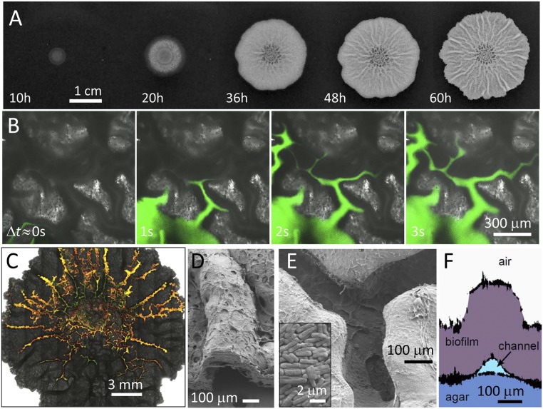
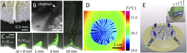
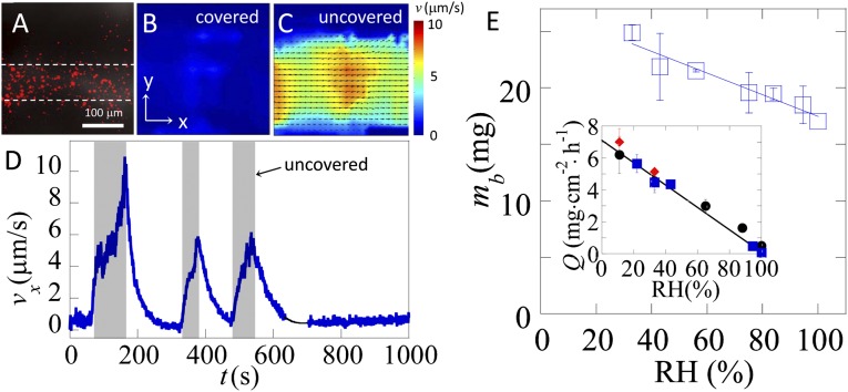
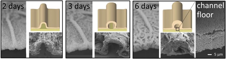
Many bacteria on earth exist in surface-attached communities known as biofilms. These films are responsible for manifold problems, including hospital-acquired infections and biofouling, but they can also be beneficial. Biofilm growth depends on the transport of nutrients and waste, for which diffusion is thought to be the main source of transport. However, diffusion is ineffective for transport over large distances and thus should limit growth. Nevertheless, biofilms can grow to be very large. Here we report the presence of a remarkable network of well-defined channels that form in wild-type Bacillus subtilis biofilms and provide a system for enhanced transport. We observe that these channels have high permeability to liquid flow and facilitate the transport of liquid through the biofilm. In addition, we find that spatial variations in evaporative flux from the surface of these biofilms provide a driving force for the flow of liquid in the channels. These channels offer a remarkably simple system for liquid transport, and their discovery provides insight into the physiology and growth of biofilms.


Conflict of interest statement

The authors declare no conflict of interest.

Figures

Fig. 1.
Fig. 1.
Characterization of channels within B. subtilis biofilms. (A) Biofilm growing on the surface of an agar gel containing water and nutrients. The biofilm increases in height to hundreds of micrometers, spreads to reach a diameter of several centimeters, and forms macroscopic wrinkles. (B) Series of microscopy images of a region near the center of the biofilm. Injection of an aqueous dye reveals a network of channels beneath the wrinkles. (C) Microscopy image of a biofilm after injection of an aqueous solution containing a mixture of fluorescent beads reveals the connectivity of the channels. (D) SEM image of a wrinkle cross-section. (E) SEM image of the underside of a biofilm reveals well-defined channels. (Inset) SEM image of the microstructure of the biofilm. (F) Side view of a biofilm wrinkle reconstructed from profiles of plastic molds of the upper and lower surfaces of the biofilm and the surface of the agar.
Fig. 2.
Fig. 2.
Reduced pressure in channels due to evaporation. (A) Photograph of an open channel. (B) Microscopy image of an air–water meniscus at the end of an open channel. (C) Series of microscopy images depicting the imbibition of fluorescent oil into an open channel. For conditions with no evaporation (pw = pw,sat), no imbibition occurs; when evaporation is induced (pw < pw,sat), the oil is imbibed far into the biofilm. (D) Infrared thermograph of a biofilm on agar exposed to ambient air with RH = 56% and T = 21.5 °C reveals spatial differences in evaporative flux. (E) Illustration of evaporation from the surface of the film (blue arrows) that drives liquid flow throughout the agar and the biofilm. (Inset) Liquid-filled channels with high permeability for liquid flow exist beneath the biofilm and facilitate the flow of liquid in the xy plane. A gradient in evaporative flux (black arrows) could drive liquid flow within the channel (white arrow).
Fig. 3.
Fig. 3.
Flow in channels driven by evaporation and correlation of evaporative flux with biofilm biomass. (A) Channel containing red fluorescent beads. (B) Velocity map of the liquid in the channel shows no net flow when the biofilm is covered (pw = pw,sat), and (C) directed flow when the biofilm is uncovered (pw < pw,sat). (D) Average velocity along the channel, vx, measured within white dashed line in A, as a function of time. Shaded regions indicate times when the biofilm is exposed to RH = 49%; unshaded regions correspond to time periods where the biofilm is covered with a small Petri dish and RH equilibrates at 100%. Black line guides the eye through a short break in the data. (E) Biofilms grown in low humidity exhibit greater biomass than those grown in high humidity. Total dry biomass mb from individual colonies grown for 60 h at 30 °C, plotted as a function of relative humidity. (Inset) Evaporative flux, Q from the surface of a biofilm and agar dish as a function of RH, measured by the total change in mass of the agar plates after 60 h. For both plots, the line is a linear fit through the data, n = 6, and error bars represent the SD.
Fig. 4.
Fig. 4.
Structural evolution of the channels. Photographs, SEM images, and illustrations depict the structural evolution of a channel over time. By 6 d the biofilm has spread to cover the floor of the channel.

References

    1. Flemming H-C, Wingender J. The biofilm matrix. Nat Rev Microbiol. 2010;8(9):623–633. - PubMed
    1. Sutherland IW. Biofilm exopolysaccharides: A strong and sticky framework. Microbiology. 2001;147(Pt 1):3–9. - PubMed
    1. Branda SS, Vik S, Friedman L, Kolter R. Biofilms: The matrix revisited. Trends Microbiol. 2005;13(1):20–26. - PubMed
    1. Ghannoum M, O’Toole GA, editors. Microbial Biofilms. Washington, DC: ASM Press; 2004.
    1. O’Toole G, Kaplan HB, Kolter R. Biofilm formation as microbial development. Annu Rev Microbiol. 2000;54:49–79. - PubMed

Publication types

LinkOut - more resources