

Ocular argyrosis secondary to long-term ingestion of silver nitrate salts
- PMID: 23271882
- PMCID: PMC3526907
- DOI: 10.2147/OPTH.S37898
Ocular argyrosis secondary to long-term ingestion of silver nitrate salts
Abstract
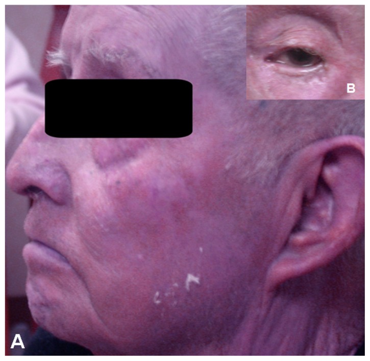
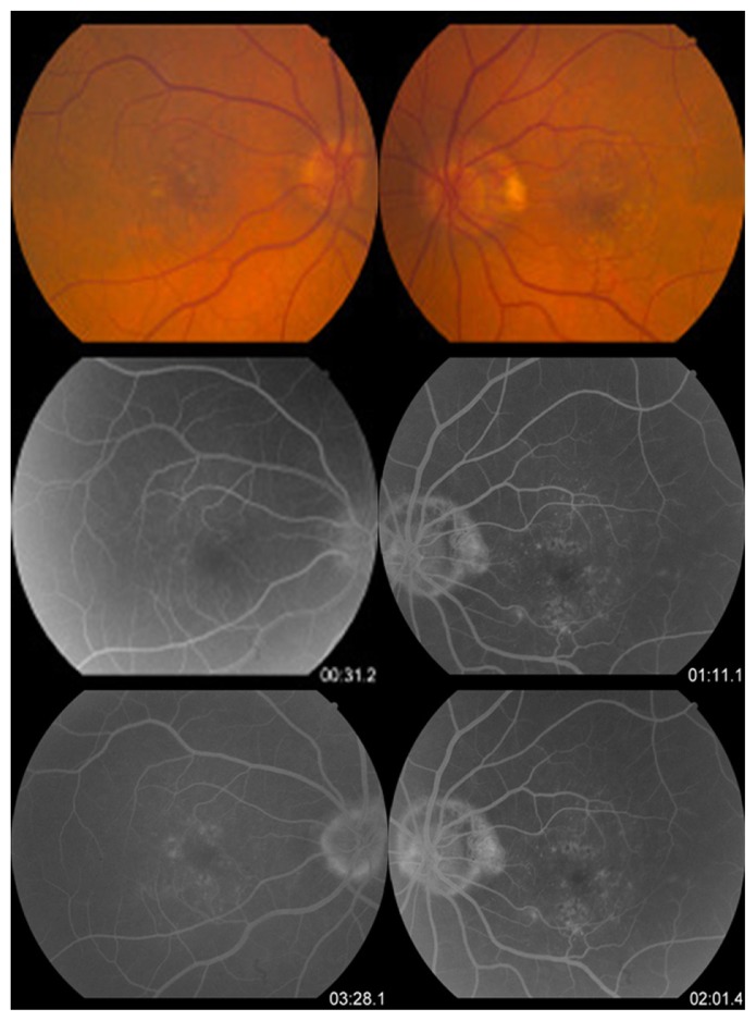
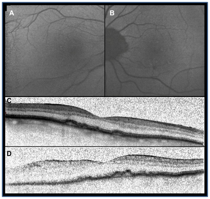
This case report describes the clinical, autofluorescence, and optical coherent tomography findings in a patient with panocular argyrosis secondary to chronic intake of diluted silver nitrate salts in his water supply. An 86-year-old Caucasian male with a distinctive gray-bluish hue of the skin presented to our clinic, having developed a slow decrease in visual acuity in both eyes and nyctalopia for the past 2 years. Based on the patient's history of chronic intake of silver nitrate salts and a positive skin biopsy (performed by the dermatology department, data not shown), a diagnosis of panocular argyrosis was made. Fluorescein angiography showed choroidal blockage with a completely dark choroid. Fundus autofluorescence was within normal limits. Optical coherent tomography showed multiple excrescences of retinal pigment epithelium in both eyes. Although the drusen-like changes on fundus examination and retinal pigment epithelium changes may account for the diminished vision, the presence of concomitant nyctalopia suggests underlying damage of the photoreceptors.
Keywords: argyria; fundus autofluorescence; ocular argyrosis; optical coherent tomography; silver nitrate.
Figures
References
-
- Alexander JW. History of the medical use of silver. Surg Infect (Larchmt) 2009;10(3):289–292. - PubMed
-
- Pala G, Fronterre A, Scafa F, et al. Ocular argyrosis in a silver craftsman. J Occup Health. 2008;50(6):521–524. - PubMed
-
- Drake PL, Hazelwood KJ. Exposure-related health effects of silver and silver compounds: a review. Ann Occup Hyg. 2005;49(7):575–585. - PubMed
-
- Sanchez-Pulgarin M, Matilla M, Martinez-de-la-Casa JM, Jerez M, Benitez-del-Castillo JM. Confocal microscopy in ocular argyrosis. Cornea. 2010;29(5):580–582. - PubMed
-
- Moss AP, Sugar A, Hargett NA, Atkin A, Wolkstein M, Rosenman KD. The ocular manifestations and functional effects of occupational argyrosis. Arch Ophthalmol. 1979;97(5):906–908. - PubMed
Publication types
LinkOut - more resources
Full Text Sources
Miscellaneous
