. 2012;8(12):e1002814.
doi: 10.1371/journal.pcbi.1002814. Epub 2012 Dec 13.

Climbing fiber burst size and olivary sub-threshold oscillations in a network setting

Affiliations

Climbing fiber burst size and olivary sub-threshold oscillations in a network setting

Jornt R De Gruijl et al. PLoS Comput Biol. 2012.

Abstract

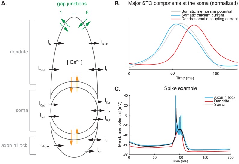
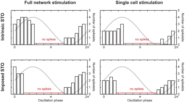
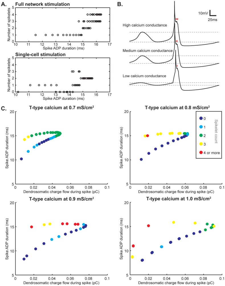
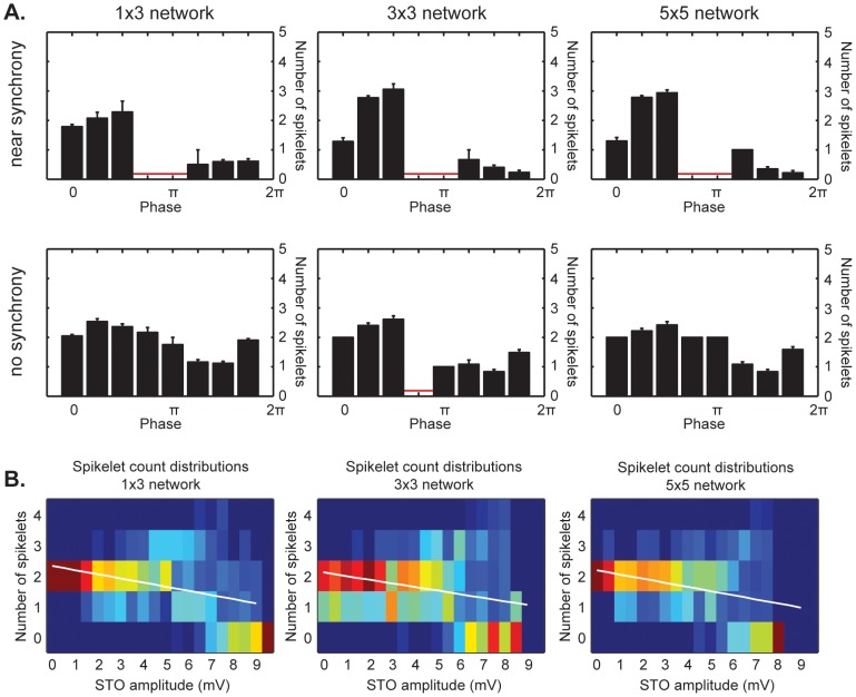
The inferior olivary nucleus provides one of the two main inputs to the cerebellum: the so-called climbing fibers. Activation of climbing fibers is generally believed to be related to timing of motor commands and/or motor learning. Climbing fiber spikes lead to large all-or-none action potentials in cerebellar Purkinje cells, overriding any other ongoing activity and silencing these cells for a brief period of time afterwards. Empirical evidence shows that the climbing fiber can transmit a short burst of spikes as a result of an olivary cell somatic spike, potentially increasing the information being transferred to the cerebellum per climbing fiber activation. Previously reported results from in vitro studies suggested that the information encoded in the climbing fiber burst is related to the occurrence of the spike relative to the ongoing sub-threshold membrane potential oscillation of the olivary cell, i.e. that the phase of the oscillation is reflected in the size of the climbing fiber burst. We used a detailed three-compartmental model of an inferior olivary cell to further investigate the possible factors determining the size of the climbing fiber burst. Our findings suggest that the phase-dependency of the burst size is present but limited and that charge flow between soma and dendrite is a major determinant of the climbing fiber burst. From our findings it follows that phenomena such as cell ensemble synchrony can have a big effect on the climbing fiber burst size through dendrodendritic gap-junctional coupling between olivary cells.


Conflict of interest statement

The authors have declared that no competing interests exist.

Figures

Figure 1
Figure 1. Architecture and electrophysiological properties of the cell model.
A. Schematic representation of the three-compartmental cell model used. From top to bottom, the compartments represent the dendrite, the soma and the axon hillock (also indicated in gray on the left). Current flows internally between the dendritic and the somatic compartment as well as between the somatic compartment and the axon hillock, as indicated in orange. In addition, current can flow between a cell and up to eight cells it is connected to through the gap junctions in the dendritic compartment, indicated in green. Each compartment has its own set of ion channels. The dendrite has a high-threshold calcium current ICaH (P/Q-type) and resultant internal calcium concentration [Ca2+], a calcium-dependent potassium current IK,Ca, a cationic current Ih and a passive leak current Ild. At the soma, there is a low-threshold calcium current ICaL (T-type), a fast sodium current INa, a potassium current with a slow component IK,s and a fast component IK,f, and a passive leak current Ils. The axon hillock compartment has a fast sodium current INa,ax, a fast potassium current IK,f and a passive leak current Ila. B. Normalized representation of the major STO components at the soma. The gray line shows the somatic membrane potential as a reference. The upward slope of the STO is caused by an activation of low-threshold calcium ion channels, leading to a depolarizing current (blue line). As the membrane potential becomes more depolarized, the calcium ion channels inactivate and current leaking from soma to dendrite increases in intensity (red line), causing the membrane potential to drop again. C. Example of a spike. A depolarizing current is applied at the dendritic compartment (red line), which exhibits a slow depolarization. The somatic compartment (black line) responds to this with a slow depolarization on top of which a fast sodium spike is generated. The axon hillock (blue line) shows fast sodium responses to the depolarization in the somatic compartment: the peak of the first sodium spike occurs before the somatic sodium spike (as reported by Mathy et al. [27]) and a burst of spikes is generated riding on the somatic depolarization. This burst of spikes is propagated back to the soma to some extent and is visible as spikelets on the calcium depolarization.
Figure 2
Figure 2. Phase dependency of AP spikelet counts.
Phase dependency of IO cell AP spikelet count under natural (intrinsic STO) conditions and when a sinusoidal 5 Hz STO is imposed through current injection. Left panels show the results for full network stimulation, corresponding with e.g. stimulating a fiber beam, whereas the right panels show the results for single-cell stimulation. Under all four conditions, the STO establishes a firing window outside of which the cell does not fire action potentials (the phase range where spikes were not generated is indicated in red), in concordance with earlier findings . However, the actual boundaries of the firing window are different when an oscillation that differs from the intrinsic STO is imposed (bottom panels as opposed to top panels). When the entire network is stimulated, there is a clear phase-dependency of AP spikelet count, as the spikelet count ranges from 1 to 4 depending on the phase (left panels). However, when only one cell in a cluster of 9 cells is stimulated, this phase dependency is less clear, as the spikelet count can still take different values, but is of limited variability and generally equals 2 (right panels).
Figure 3
Figure 3. Factors underlying AP spikelet count.
A. Correlation between spike ADP duration and AP spikelet count. In general, a longer ADP increases the chances of larger amounts of spikelets on top of the somatic ADP. This is most readily apparent when stimulating the entire network (top panel). Still, the ADP duration by itself does not provide an adequate explanation for the amount of spikelets that are part of the spike shape, since the number of spikelets can still vary considerably even within a millisecond bin both when the entire networks fires and when only one cell does. When only one cell in the network fires, the relation between ADP duration and number of spikelets does not appear to be linear, even though on average a higher number of spikelets is still more likely at longer ADP durations (bottom panel). B. Changing the intrinsic conductance values of the low-threshold calcium current changes the amplitude of the oscillations (as indicated by the dashed lines aligned with the peaks and troughs of the depicted traces), but also the number of spikelets (time of occurrence is indicated with red markers in each depicted trace). The number of spikelets decreases as the T-type calcium expression level decreases. C. Spike ADP and dendrosomatic coupling currents as AP spikelet count determinants. Single cells in a 9-cell network were stimulated for all of the simulation results shown. Warmer colors represent higher numbers of AP spikelets (color-coding is the same for all four panels and indicated in the figure). Spike ADP duration and dendrosomatic charge flow form a trajectory. It is readily apparent from all four panels that higher numbers of spikelets are more likely to occur at longer ADPs, but in addition decreased charge flow increases the chance of generating an AP with more spikelets. As a result, the prediction of AP spikelet count can be improved when taking both ADP duration and dendrosomatic charge flow into account. At different T-type calcium expression levels, the range of possible spikelet counts and the distributions thereof vary. Clearly, spike ADP and dendrosomatic coupling currents are major determinants for the AP spikelet count measured at the soma, but other currents both intra- and intercellular can cause local phenomena in the distributions along the trajectories shown.
Figure 4
Figure 4. Effects of network synchrony on AP spikelet count.
A. Phase dependency of AP spikelet count for different network sizes when a network consists of coupled cells with near-synchronized STOs (STO amplitude >5.5 mV, top row) or non-synchronized STOs (STO amplitude <5.5 mV, bottom row). Phase ranges where no spikes were fired are indicated in red. Top row, near synchrony - The small 1×3 network shows a weak phase dependency of the AP spikelet count, averaging 0.5 spikelets on the upward slope of the STO and 2 spikelets around the peak. The 3×3 and 5×5 networks show similar outcomes and a stronger phase-dependency of the AP spikelet count, averaging 0 spikelets on the upward slope, followed by 1 spikelet near the peak and 3 spikelets around the peak. Due to a strong depolarizing current (5 pA, 20 ms for 1×3 networks and 6 pA, 20 ms for 3×3 and 5×5 networks, see Methods), spikes are sometimes fired outside the usual firing window in the trough of the oscillation (1.25π radians bin, total of 7 occurrences in 2100 simulations across all network sizes). Bottom row, no synchrony - Regardless of network size, there is no clear phase-dependency of AP spikelet count. A rounded average of 2 spikelets is seen across all phases, except for part of the phase range falling outside or close to the bounds of the firing window where a rounded average of 1 spikelet may occur. Due to analysis restrictions imposed by determining the phase, STO amplitudes smaller than 1 mV are poorly represented in the data set. B. AP spikelet distributions across STO amplitudes for different network sizes. Warmer colors denote more occurrences, cooler colors less. The distribution of spikelets changes as a function of STO amplitude. In a range of approximately 5 to 7 mV, the distribution broadens, corresponding with the phase dependency of AP spikelet counts shown in panel A. At an STO amplitude of 7.5 mV or more, this distribution narrows again, but at a lower average spikelet count than was seen at lower amplitudes. The average number of AP spikelets shows a downward trend for all network sizes, as illustrated by the fitted trend lines shown in white.

Similar articles

Cited by

References

    1. De Zeeuw CI, Hoebeek FE, Bosman LW, Schonewille M, Witter L, et al. (2011) Spatiotemporal firing patterns in the cerebellum. Nat Rev Neurosci 12(6): 327–44. - PubMed
    1. Eccles J, Llinás R, Sasaki K (1964) Excitation of cerebellar Purkinje cells by the climbing fibres. Nature 203: 245–246. - PubMed
    1. Deuschl G, Toro C, Valls-Solé J, Zeffiro T, Zee DS, et al. (1994) Symptomatic and essential palatal tremor. 1. Clinical, physiological and MRI analysis. Brain 117: 775–788. - PubMed
    1. Holmberg M, Duyckaerts C, Dürr A, Cancel G, Gourfinkel-An I, et al. (1998) Spinocerebellar ataxia type 7 (SCA7): a neurodegenerative disorder with neuronal intranuclear inclusions. Hum Mol Genet 7: 913–918. - PubMed
    1. Raike RS, Jinnah HA, Hess EJ (2005) Animal models of generalized dystonia. NeuroRx 2: 504–512. - PMC - PubMed

Publication types