. 2012;8(12):e1003065.
doi: 10.1371/journal.ppat.1003065. Epub 2012 Dec 13.

Antigenic subversion: a novel mechanism of host immune evasion by Ebola virus

Affiliations

Antigenic subversion: a novel mechanism of host immune evasion by Ebola virus

Gopi S Mohan et al. PLoS Pathog. 2012.

Abstract

In addition to its surface glycoprotein (GP(1,2)), Ebola virus (EBOV) directs the production of large quantities of a truncated glycoprotein isoform (sGP) that is secreted into the extracellular space. The generation of secreted antigens has been studied in several viruses and suggested as a mechanism of host immune evasion through absorption of antibodies and interference with antibody-mediated clearance. However such a role has not been conclusively determined for the Ebola virus sGP. In this study, we immunized mice with DNA constructs expressing GP(1,2) and/or sGP, and demonstrate that sGP can efficiently compete for anti-GP(12) antibodies, but only from mice that have been immunized by sGP. We term this phenomenon "antigenic subversion", and propose a model whereby sGP redirects the host antibody response to focus on epitopes which it shares with membrane-bound GP(1,2), thereby allowing it to absorb anti-GP(1,2) antibodies. Unexpectedly, we found that sGP can also subvert a previously immunized host's anti-GP(1,2) response resulting in strong cross-reactivity with sGP. This finding is particularly relevant to EBOV vaccinology since it underscores the importance of eliciting robust immunity that is sufficient to rapidly clear an infection before antigenic subversion can occur. Antigenic subversion represents a novel virus escape strategy that likely helps EBOV evade host immunity, and may represent an important obstacle to EBOV vaccine design.


Conflict of interest statement

The authors have declared that no competing interests exist.

Figures

Figure 1
Figure 1. Diagram of EBOV RNA editing and construction of EBOV GP mutants.
(A) Schematic diagram of GP1,2 and sGP. Membrane-bound GP1,2 is encoded in the EBOV genome in two disjointed reading frames. The GP editing site is a tract of 7 A's approximately 900 nucleotides downstream of the start codon. Slippage of EBOV RNA-dependent RNA polymerase at the editing site results in insertion of an 8th-A which brings the two GP reading frames in register resulting in read-through translation of full-length membrane-bound trimeric GP1,2. Unedited transcripts contain a premature stop codon and produce truncated dimerized sGP. (B) EBOV GP and editing site mutants. Mutated nucleotides are shown in red and the primary gene products expressed by these constructs are also listed. (C) Expression of EBOV GP by wild type and mutant DNA constructs. HeLa cells were transfected with the wild type GP or editing site mutant constructs and GP expression was assayed by Western blot at 48 h post-transfection.
Figure 2
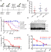
Figure 2. Immunogenicity of EBOV GP editing site mutants.
(A) Immunization study design. Female BALB/C mice were immunized with the four editing site mutant constructs in the pCAGGS vector. Mice were vaccinated IM with 50 µg of DNA (25 µg/leg) according to the schedule shown. (B) Antibody response against GP1,2. (C) Antibody response against sGP. The levels of antibody response induced by EBOV GP DNA constructs in mice were measured by ELISA using His-GP1,2 or His-sGP as coating antigen. Antibody concentration was determined from a standard curve and expressed as µg/mL of anti-GP IgG. Asterisks indicate statistically significant difference between groups and P-values are given in red. (D) Comparison of antibody levels against GP1,2 and sGP induced by each EBOV GP DNA construct. Average titers of anti-GP1,2 (blue) and anti-sGP (red) antibodies within immunization groups are shown for comparison of the GP isoform reactivity profiles both within and between immunization groups. Asterisks indicate statistically significant differences between anti-GP1,2 and anti-sGP titers within groups, as measured by paired, two-tailed Student's t-test (* = p<0.05, ** = p<0.001).
Figure 3
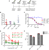
Figure 3. Antiserum from mice immunized against GP1,2 or sGP display different reactivity patterns.
(A) Detection by Western blot of antibodies against GP1,2 and sGP from immunized mice. 50 ng of purified His-sGP and His-GP1,2 were run by SDS-PAGE under denaturing conditions and probed with 1∶1000 pooled GP1,2Edit or sGPEdit antisera followed by blotting with HRP-conjugated goat anti-mouse IgG. (B) Schematic of competition ELISA. Wells were coated with GP1,2 and incubated with pooled antisera as well as increasing concentrations of competing antigen (sGP or GP1,2) to compete for antibodies. After two hours, plates were washed and then incubated with HRP-conjugated secondary antibody followed by addition of substrate to develop color. (C, D) Competition ELISA. Antisera from mice immunized with sGPEdit, GP-7A, GP-8A, and GP1,2Edit were diluted to give similar anti-GP1,2 signal. Diluted antiserum was mixed with increasing quantities of purified His-sGP (C) or His-GP1,2 (D) and incubated in His-GP1,2 coated wells and developed as described above. Experiments were performed in duplicate and repeated at least three times, with representative results shown. (E, F) Competition Immunoprecipitation. Pooled antisera from GP1,2Edit-immunized mice (E) or sGP-immunized mice (F) were incubated with no GP, purified sGP or GP1,2 alone, or with fixed GP1,2 and increasing concentrations of sGP to compete for anti-GP1,2 antibodies. GP1,2 was incubated with recombinant HA as a negative control. The upper panel for the sGPEdit antisera shows the GP1,2 portion of the blot at a longer exposure time to show the attenuation of signal with increasing sGP concentration. Results are representative of three independent experiments.
Figure 4
Figure 4. Interference with antibody-dependent neutralization by sGP.
(A) Neutralization of EBOV GP pseudovirus. Neutralizing activity of antisera was determined by incubating 500 pfu of GP1,2-pseudotyped virus with dilutions of pooled GP1,2-immunized (Blue), sGP-immunized (Red), and empty pCAGGS vector-immunized (black) antisera. Neutralization was measured as decrease in luciferase expression compared to virus-only controls after 48 h. (B) Interference of EBOV GP pseudovirus neutralization by sGP. The ability of sGP to interfere with antibody-dependent neutralization was determined by allowing sGP to compete with GP1,2 pseudotyped viruses for anti-GP1,2 antibodies. Pooled GP1,2-immunized (blue) and sGP-immunized (red) antisera were fixed at the dilution corresponding to 80% neutralization. Antisera was co-incubated with increasing dilutions of His-tagged sGP (solid markers) or His-tagged influenza PR8 HA (open markers), and rescue of infectivity was measured as described in methods.
Figure 5
Figure 5. Comparison of binding affinity of GP1,2-immunized versus sGP-immunized antisera for sGP and GP1,2.
(A) Determining apparent Kd value of antibodies from immunized mice for GP1,2 and sGP. Antiserum from five mice immunized against GP1,2 and five mice immunized against sGP were individually analyzed by quantitative ELISA using GP1,2 (blue) or sGP (red) as coating antigen. Scatchard analysis was used to calculate apparent dissociation constants (Kd). (B) Comparison of antibody affinity for GP1,2 and sGP. Comparison of apparent Kd's of GP1,2-immunized and sGP-immunized polyclonal antisera for sGP (red) and GP1,2(blue) was determined by nonlinear regression analysis of Scatchard plots. Kd's for sGP and GP1,2 were calculated for five individual mice in each group and values for the same animal are connected by a black line.
Figure 6
Figure 6. The effect of sGP on immune response when antigen exposure mimics natural infection.
(A) Immunization study design. Female BALB/C mice were immunized IM with 50 µg of total DNA per immunization according to the schedule shown. Mice were immunized with a 3∶1 ratio of sGP Edit∶GP1,2 Edit in pCAGGS. Control groups were immunized with sGP Edit or GP1,2 Edit alone plus empty pCAGGS vector to keep total amount of immunizing DNA constant. (B) Comparison of antibody response against GP1,2. Mouse sera collected at week 6 were analyzed for anti-GP1,2 antibodies by ELISA using GP1,2 as coating antigen. (C) sGP competition ELISA. The ability of sGP to compete for anti-GP antibodies was determined by competition ELISA as in Figure 3B. Pooled antisera were analyzed from mice immunized with a GP1,2 Edit (blue), sGP Edit (red), or a 3∶1 ratio of sGP Edit∶GP1,2Edit (purple), and were diluted to give roughly equivalent anti-GP1,2 signal. Competition ELISA was performed from antisera collected at both week 6 (light color) and week 12 (dark color) according to the immunization schedule. (D) Competition immunoprecipitation. Pooled antisera from sGPEdit+GP1,2Edit-immunized mice were incubated with no GP, purified sGP or GP1,2 alone, or with fixed GP1,2 and increasing concentrations of sGP to compete for anti-GP1,2 antibodies. GP1,2 was incubated with recombinant HA as a negative control, and precipitated and analyzed as in Figure 3E,F. (E) Neutralization of EBOV GP pseudovirus. Neutralizing activity of antisera was determined by incubating 500 pfu of GP1,2-pseudotyped virus with dilutions of pooled sGP+GP1,2-immunized (red), or empty pCAGGS vector-immunized (black) antisera. Neutralization was measured as decrease in luciferase expression compared to virus-only controls. (F) Interference of EBOV GP pseudovirus neutralization by sGP. The ability of sGP to interfere with antibody-dependent neutralization was determined as in Figure 4B. Pooled sGP+GP1,2-immunized antisera were fixed at the dilution corresponding to 80% neutralization. Antisera were co-incubated with increasing dilutions of purified sGP (red) or purified influenza PR8 HA (blue), and rescue of infectivity was measured as described in methods.
Figure 7
Figure 7. Ability of sGP to divert antibody responses against GP1,2.
(A) Immunization study design. Female BALB/C mice were immunized IM with 50 µg of total DNA per immunization according to the schedule. Two groups of mice (n = 12) were primed and boosted as in previous experiments with either sGP Edit or GP1,2 Edit in pCAGGS vector. Each group was divided in two and subgroups were boosted at week 10 with either the same construct against which they had initially been immunized, or with the opposite editing site mutant construct. (B) Comparison of antibody response against GP1,2. Sera collected at week 12 were analyzed for antibodies against GP1,2 by ELISA using GP1,2 as coating antigen. (C) sGP competition ELISA. The ability of sGP to compete for anti-GP1,2 antibodies was determined by competition ELISA as described in Figure 3B. Pooled antisera were analyzed from mice immunized with sGP Edit and then boosted at week 10 with either GP1,2 Edit (red), or sGP Edit (purple), and from mice immunized with GP1,2 Edit and then boosted at week 10 with either GP1,2Edit (blue) or sGP Edit (green). All ELISA experiments were performed in duplicate at least three times and representative results shown. (D) Interference of EBOV GP pseudovirus neutralization by sGP. The ability of sGP to interfere with antibody-dependent neutralization was determined as in Figure 4B. Pooled sGP-primed, GP1,2-boosted (red) and GP1,2-primed, sGP-boosted (green) antisera were fixed at the dilution corresponding to 50% neutralization. Antisera were co-incubated with increasing dilutions of His-tagged sGP (solid markers) or His-tagged influenza PR8 HA (open markers), and rescue of infectivity was measured as described in methods. (E) Comparison of 50% neutralization titers. Antiserum titers corresponding to 50% pseudovirus neutralization activity (NT50) were calculated for week 6 (fine checkered) and week 12 (coarse checkered) mice. Error bars correspond to 95% confidence interval as determined by Student's t-test.
Figure 8
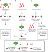
Figure 8. Proposed mechanism for antigenic subversion.
Regions of GP1,2 that are shared with sGP are in red, while unshared epitopes are in green. B-cells are colored according to the regions of GP1,2 and sGP against which they react. (A) A naïve animal begins with B-cells that can potentially recognize epitopes distributed throughout GP1,2 and sGP. When sGP is expressed at much higher levels than GP1,2, as occurs during infection, those B-cells that recognize sGP epitopes, many of which are shared with GP1,2 (red regions of sGP and GP1,2) are preferentially activated and expanded compared to B-cells that recognize unshared epitopes of GP1,2 (green regions of GP1,2). Thus, sGP-reactive antibodies dominate the immune response. (B) Prior immunization by sGP. Because sGP shares over 90% of its linear sequence with GP1,2, animals primed with sGP generate anti-sGP antibodies, many of which are directed against epitopes shared with GP1,2. When these animals (or individuals who have previously been infected and recovered from EBOV infection) are boosted with GP1,2, sGP cross-reactive memory cells outnumber and express higher affinity receptors than naïve GP1,2 specific B-cells, resulting in preferential expansion of these sGP-cross-reactive B-cells and a predominantly sGP-reactive immune response. (C) Prior immunization by GP1,2. Priming naïve animals with GP1,2 results in antibodies largely against GP1,2 epitopes not shared with sGP, presumably due to the immunodominance and high accessibility of the GP1,2 mucin domain and shielding of shared epitopes. When these animals are boosted with sGP, or if they are infected with EBOV and do not have sufficiently high titers of anti-GP1,2 antibodies to clear the infection rapidly, memory B-cells that recognize shared epitopes encounter their cognate antigen and expand, while non-cross-reactive GP1,2-specific B-cells are not boosted, resulting in subversion of the host immune response towards sGP cross-reactivity. (D) Successful clearance of EBOV infection. In order to avoid sGP-mediated antigenic subversion, high enough titers of non-crossreactive anti-GP1,2 antibodies must be maintained to rapidly clear EBOV infection before subversion can occur.

Comment in

References

    1. Feldmann H, Geisbert TW (2011) Ebola haemorrhagic fever. Lancet 377: 849–862. - PMC - PubMed
    1. Belyi VA, Levine AJ, Skalka AM (2010) Unexpected inheritance: multiple integrations of ancient bornavirus and ebolavirus/marburgvirus sequences in vertebrate genomes. PLoS Pathog 6: e1001030. - PMC - PubMed
    1. Taylor DJ, Leach RW, Bruenn J (2010) Filoviruses are ancient and integrated into mammalian genomes. BMC Evol Biol 10: 193. - PMC - PubMed
    1. Hayman DT, Emmerich P, Yu M, Wang LF, Suu-Ire R, et al. (2010) Long-term survival of an urban fruit bat seropositive for Ebola and Lagos bat viruses. PLoS One 5: e11978. - PMC - PubMed
    1. Leroy EM, Epelboin A, Mondonge V, Pourrut X, Gonzalez JP, et al. (2009) Human Ebola outbreak resulting from direct exposure to fruit bats in Luebo, Democratic Republic of Congo, 2007. Vector Borne Zoonotic Dis 9: 723–728. - PubMed

Publication types

MeSH terms