. 2012;8(12):e1003119.
doi: 10.1371/journal.pgen.1003119. Epub 2012 Dec 13.

A mechanism of gene amplification driven by small DNA fragments

Affiliations

A mechanism of gene amplification driven by small DNA fragments

Kuntal Mukherjee et al. PLoS Genet. 2012.

Abstract

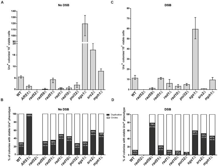
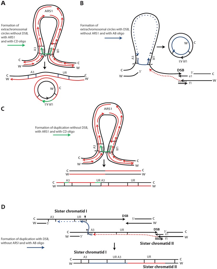
DNA amplification is a molecular process that increases the copy number of a chromosomal tract and often causes elevated expression of the amplified gene(s). Although gene amplification is frequently observed in cancer and other degenerative disorders, the molecular mechanisms involved in the process of DNA copy number increase remain largely unknown. We hypothesized that small DNA fragments could be the trigger of DNA amplification events. Following our findings that small fragments of DNA in the form of DNA oligonucleotides can be highly recombinogenic, we have developed a system in the yeast Saccharomyces cerevisiae to capture events of chromosomal DNA amplification initiated by small DNA fragments. Here we demonstrate that small DNAs can amplify a chromosomal region, generating either tandem duplications or acentric extrachromosomal DNA circles. Small fragment-driven DNA amplification (SFDA) occurs with a frequency that increases with the length of homology between the small DNAs and the target chromosomal regions. SFDA events are triggered even by small single-stranded molecules with as little as 20-nt homology with the genomic target. A double-strand break (DSB) external to the chromosomal amplicon region stimulates the amplification event up to a factor of 20 and favors formation of extrachromosomal circles. SFDA is dependent on Rad52 and Rad59, partially dependent on Rad1, Rad10, and Pol32, and independent of Rad51, suggesting a single-strand annealing mechanism. Our results reveal a novel molecular model for gene amplification, in which small DNA fragments drive DNA amplification and define the boundaries of the amplicon region. As DNA fragments are frequently found both inside cells and in the extracellular environment, such as the serum of patients with cancer or other degenerative disorders, we propose that SFDA may be a common mechanism for DNA amplification in cancer cells, as well as a more general cause of DNA copy number variation in nature.


Conflict of interest statement

The authors have declared that no competing interests exist.

Figures

Figure 1
Figure 1. Schemes of different strains containing the A3-UR amplicon cassette in chromosome VII.
The yeast strain FRO-155 has a GSHU cassette in the middle of the TRP5 gene on chromosome VII. (C) and (W) indicate the Crick and Watson strands. The yeast/E. coli shuttle plasmid YRpKM1, containing the AmpR gene (dark blue), ORI (yellow), the yeast ARS1 (blue) and the yeast URA3 marker gene (red), was used to generate the amplicon cassette. In order to integrate the amplicon cassette into yeast chromosomal DNA, YRpKM1 was linearized by the NcoI (N) enzyme in the middle of the URA3 gene. The linearized YRpKM1, A3-UR cassette, was integrated into S. cerevisiae chromosome VII at the site of an I-SceI DSB within the TRP5 locus of strain FRO-155 following co-transformation with two pairs of oligos (Trp5.A3.F, Trp5.A3.R/Trp5.UR.F, Trp5.UR.R, see Table S1) complementary to the ends of the A3-UR cassette and to the TRP5 broken ends, according to the gene collage protocol, generating strains KM193 and KM196. Correct integration of the A3-UR amplicon cassette was confirmed by PCR and sequence analysis. Yeast strains KM-201,203 and KM-209,211 derive from KM-193 and KM-196, respectively. KM-201,203 strain contain a LEU2 marker (light blue) integrated downstream of the UR sequence. In strain KM-209,211 the UR sequence is replaced by the LEU2 gene. The KM-221,222 and KM-257,259 strains are variant forms of KM-201,203 containing the GSH cassette with the I-SceI endonuclease gene under the inducible GAL1 promoter, the hygromycin resistance marker and the I-SceI cutting site, which was integrated 10 kb downstream or upstream from the A3-UR amplicon cassette, respectively. The KM-347,349 strains are variant forms of KM201/203 in which ARS1 has been replaced with kanMX4. The XbaI (X) and the SacI (S) restriction sites present on the chromosomal tracts without and with the amplicon are indicated.
Figure 2
Figure 2. Diagrams of SFDA products.
Schemes of the region containing the A3-UR amplicon cassette on chromosome VII in strains KM201/203 (No DSB system) and KM221/222 (DSB system) are shown. The AB (in blue) and CD (in green) oligos (not to scale), used to initiate the SFDA events, are complementary to each other and complementary to the C or W strand, respectively. The thick dotted grey line connecting the AB or CD oligo halves indicate that the two halves of the oligos are not separated from each other. The SacI (S) and XbaI (X) restriction enzyme sites present on the DNA molecules containing the amplicon and on the AB and CD oligos are indicated. If more than one site for SacI or XbaI is present on the same molecule of the amplicon these sites are identified by a small number. The base-pair sizes of all the SacI or XbaI digestion products of the regions containing the amplicon cassette are shown. The products of SFDA as intact chromosomal amplicon plus extrachromosomal circle or as tandem duplication of the amplicon cassette on the chromosome are shown.
Figure 3
Figure 3. SFDA efficiency without and with a DSB.
Presented are numbers of Ura+ colonies per 107 viable cells obtained after transformation of yeast cells with no oligos, ABS and/or CDS oligos. The vertical bars correspond to the median values from six determinations; the error bars represent the range. (A) Strains KM-201,203; (B) strains KM-221,222, in which a DSB was induced 10 kb downstream from the amplicon cassette prior to oligo transformation; and (C) strains KM-257,259, in which a DSB was induced 10 kb upstream of the amplicon cassette prior to oligo transformation. Frequency values obtained for the single-stranded ABS and CDS oligos in a given strain background were compared with each other by the Mann-Whitney test and the p values of the significant differences, highlighted by the asterisks, are given on top of the corresponding bars.
Figure 4
Figure 4. Phenotypic and molecular characterization of SFDA.
(A) Ura+ colonies from KM-221 on Ura medium after DSB induction and transformation by ABS+CDS. Arrows point to large colonies. (B) Ura+-stability test for two large and two small Ura+ colonies from strain KM-221 after DSB induction and transformation by the ABS+CDS oligos. (C) Detection of the amplicon region from large and small Ura+ colonies derived after DSB induction and transformation by the ABX and CDX oligos. Lane 1, control YRpKM1 linearized (7,045-bp); lane 2, control genomic DNA from KM-221 Ura cells (12,276-bp); lanes 3, 4 genomic DNA from large Ura+ KM-221 colonies (7,449-bp, 11,872-bp); lanes 5, 6 genomic DNA from small Ura+ KM-221 colonies (7,045-bp, 12,276-bp). (D) Detection of the amplicon region from large and small Ura+ colonies derived after DSB induction and transformation by the ABS and CDS oligos. Lanes 1, 2, genomic DNA from small Ura+ KM-221 colonies grown 1 day in Ura medium (7,045-bp, 24,514-bp); lanes 3, 4, genomic DNA from large Ura+ KM-221 colonies grown 1 day in Ura medium (8,597-bp, 22,962-bp); lanes 5, 6, genomic DNA from small Ura+ KM-221 colonies grown 7 days in Ura medium (7,045-bp, 8,597-bp, 22,962-bp); lanes 7, 8, genomic DNA from large Ura+ KM-221 colonies grown 7 days in Ura medium (8,597-bp, 22,962-bp); lane 9, control genomic DNA from KM-221 Ura cells (23,203-bp); lane 10, control YRpKM1 linearized (7,045-bp). (E) Percentage of colonies with stable Ura+ phenotype (indicative of duplication) following transformation by ABS and/or CDS oligos without DSB and with DSB at day 1 and day 7 (median and range). (F) Ura+-stability test for the large and small Ura+ colonies analyzed in (D). Single-colony streaks or dilutions on YPD and results of replica plating on Ura medium for colonies taken at day 1 or 7 of growth in the Ura medium are shown.
Figure 5
Figure 5. Genetic requirements for SFDA.
Presented are numbers of Ura+ colonies per 107 viable cells obtained after transformation by ABS and CDS oligos. The vertical bars correspond to the median values from at least six independent transformations; the error bars represent the range. (A) Strains used were KM-201,203 and derivative mutants. (C) Strains used were KM-221,222 and derivative mutants. (B) and (D) Percentage (median and range) of colonies with stable Ura+ phenotype (indicative of duplication) within four samples of 45 Ura+ colonies for each strain from the experiment shown in (A) or (C), respectively. For the percentage data shown in (B) for rad59Δ three groups of 12, 13, 12 Ura+ colonies and in (D) for sgs1Δ eight groups of 45 Ura+ colonies and for rad59Δ three groups of 33, 35, 68 colonies were screened. For the ARS1Δ strains all groups had 100% stable Ura+ colonies.
Figure 6
Figure 6. Possible SFDA mechanisms.
Small DNA fragments can find homology with single-stranded sequences at the boundaries of the A3-UR amplicon region, either during DNA replication or during processing of DSB ends, and trigger amplification events resulting in formation of extrachromosomal circles or duplications. There are multiple possible mechanisms for SFDA and here we present the sketch of four SFDA events initiated by either the AB or CD oligo. Our models aim to show examples of SFDA-driven events illustrating an intermediate step for each chosen SFDA event in which the oligos are fully paired with the chromosomal sequence. (A) SFDA-driven formation of an extrachromosomal circle by the CD oligo (in green) when no DSB is induced and there is ARS1 in the amplicon, or (B) by the AB oligo (in blue) when a DSB is induced next to the amplicon; (C) SFDA-driven formation of a tandem duplication by the CD oligo when no DSB is induced and there is ARS1 in the amplicon, or (D) by the AB oligo when a DSB is induced next to the amplicon. DNA synthesis on the Crick (C) and Watson (W) strands is indicated by the red lines, assuming the fork comes from ARS1 in (A) and (C). DSB repair synthesis is indicated as red dotted lines in (B) and (D). DNA synthesis primed by the AB oligo is shown as blue dotted line. The small black arrows indicate points of strand cleavage to resolve the recombination intermediate.

Similar articles

Cited by

References

    1. Albertson DG (2006) Gene amplification in cancer. Trends Genet 22: 447–455. - PubMed
    1. Szerlip NJ, Pedraza A, Chakravarty D, Azim M, McGuire J, et al. (2012) Intratumoral heterogeneity of receptor tyrosine kinases EGFR and PDGFRA amplification in glioblastoma defines subpopulations with distinct growth factor response. Proc Natl Acad Sci U S A 109: 3041–3046. - PMC - PubMed
    1. Albertson DG, Collins C, McCormick F, Gray JW (2003) Chromosome aberrations in solid tumors. Nat Genet 34: 369–376. - PubMed
    1. Schimke RT (1984) Gene amplification, drug resistance, and cancer. Cancer Res 44: 1735–1742. - PubMed
    1. Hattinger CM, Stoico G, Michelacci F, Pasello M, Scionti I, et al. (2009) Mechanisms of gene amplification and evidence of coamplification in drug-resistant human osteosarcoma cell lines. Genes Chromosomes Cancer 48: 289–309. - PubMed

Publication types

MeSH terms

Substances