. 2012;8(12):e1003149.
doi: 10.1371/journal.pgen.1003149. Epub 2012 Dec 13.

Base damage within single-strand DNA underlies in vivo hypermutability induced by a ubiquitous environmental agent

Affiliations

Base damage within single-strand DNA underlies in vivo hypermutability induced by a ubiquitous environmental agent

Kin Chan et al. PLoS Genet. 2012.

Abstract

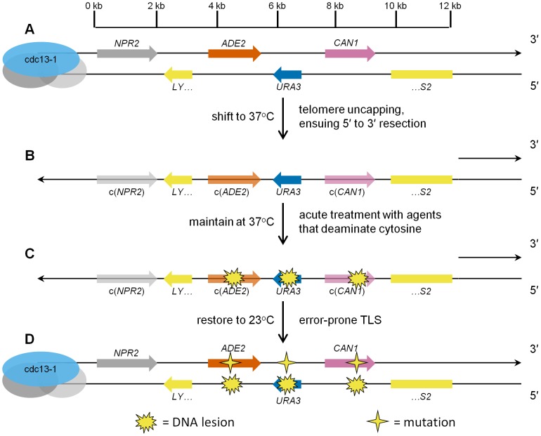
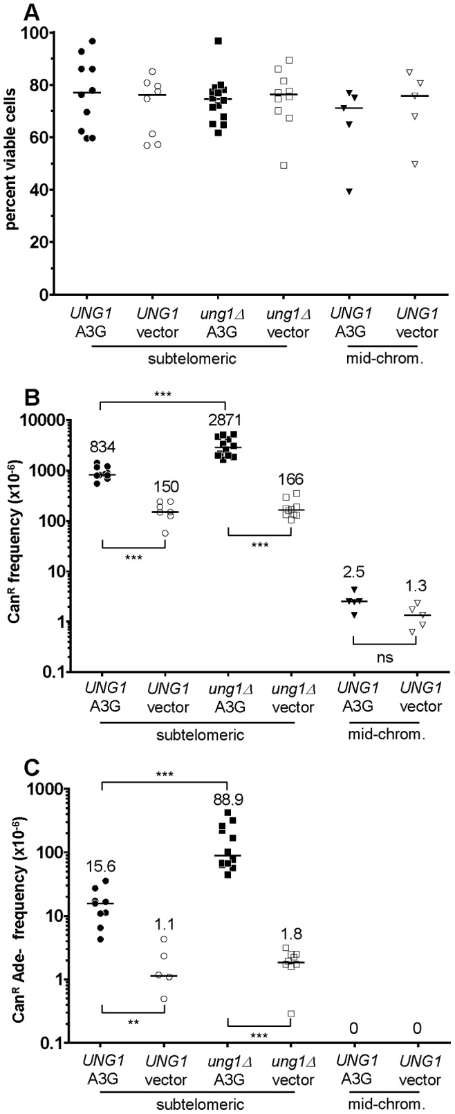
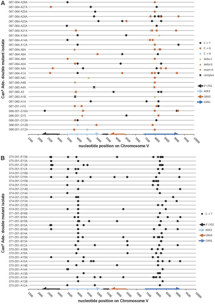
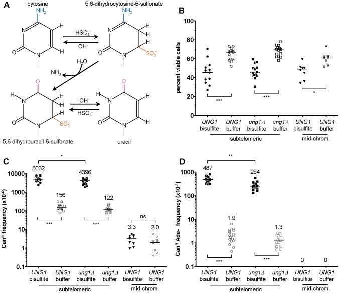
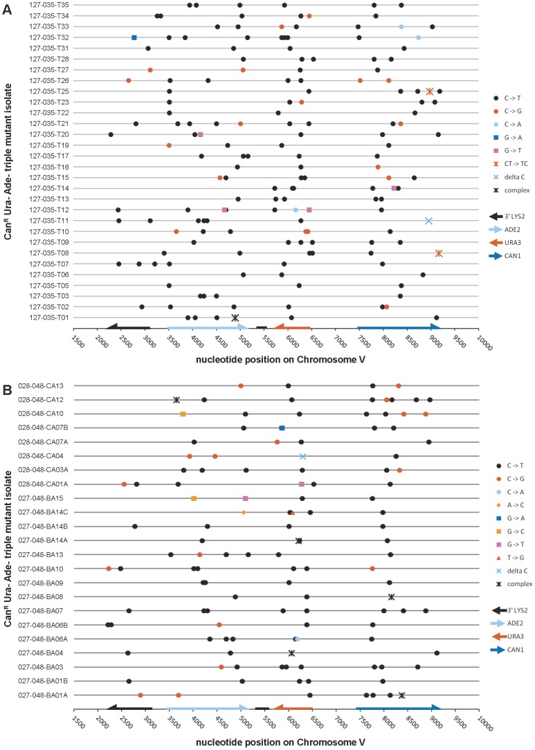
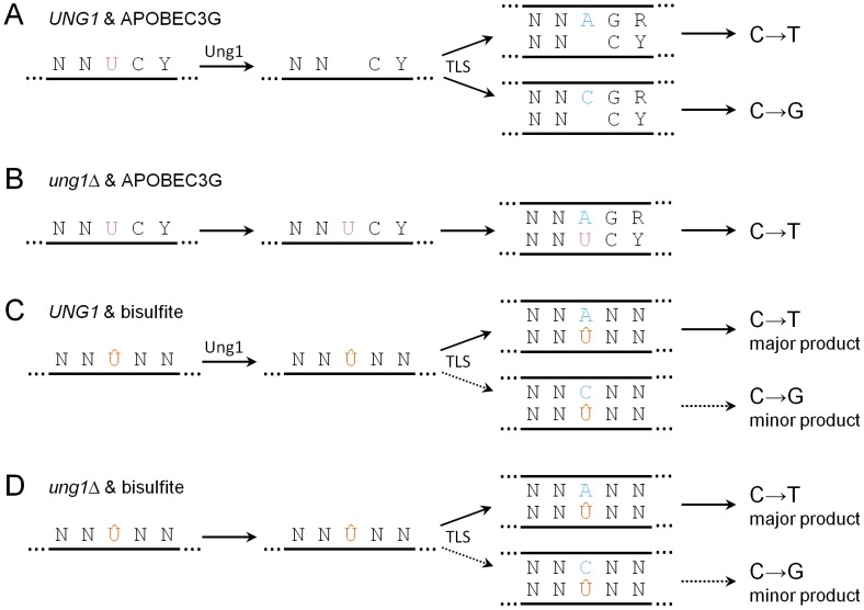
Chromosomal DNA must be in single-strand form for important transactions such as replication, transcription, and recombination to occur. The single-strand DNA (ssDNA) is more prone to damage than double-strand DNA (dsDNA), due to greater exposure of chemically reactive moieties in the nitrogenous bases. Thus, there can be agents that damage regions of ssDNA in vivo while being inert toward dsDNA. To assess the potential hazard posed by such agents, we devised an ssDNA-specific mutagenesis reporter system in budding yeast. The reporter strains bear the cdc13-1 temperature-sensitive mutation, such that shifting to 37°C results in telomere uncapping and ensuing 5' to 3' enzymatic resection. This exposes the reporter region, containing three closely-spaced reporter genes, as a long 3' ssDNA overhang. We validated the ability of the system to detect mutagenic damage within ssDNA by expressing a modified human single-strand specific cytosine deaminase, APOBEC3G. APOBEC3G induced a high density of substitutions at cytosines in the ssDNA overhang strand, resulting in frequent, simultaneous inactivation of two reporter genes. We then examined the mutagenicity of sulfites, a class of reactive sulfur oxides to which humans are exposed frequently via respiration and food intake. Sulfites, at a concentration similar to that found in some foods, induced a high density of mutations, almost always as substitutions at cytosines in the ssDNA overhang strand, resulting in simultaneous inactivation of at least two reporter genes. Furthermore, sulfites formed a long-lived adducted 2'-deoxyuracil intermediate in DNA that was resistant to excision by uracil-DNA N-glycosylase. This intermediate was bypassed by error-prone translesion DNA synthesis, frequently involving Pol ζ, during repair synthesis. Our results suggest that sulfite-induced lesions in DNA can be particularly deleterious, since cells might not possess the means to repair or bypass such lesions accurately.


Conflict of interest statement

The authors have declared that no competing interests exist.

Figures

Figure 1
Figure 1. Schematic representation of ssDNA mutagenesis reporter system.
(A) Three reporter genes, ADE2, URA3, and CAN1, were relocated from their respective native genomic loci into the subtelomeric region of Chromosome V, within a haploid budding yeast strain bearing the temperature-sensitive cdc13-1 mutation, thus creating strain ySR127. The 0 kb mark in the scale bar denotes the start of unique DNA sequence (conversely, the end of telomeric repeat sequences). (B) Shifting ySR127 cells to 37°C results in telomere uncapping. Subsequent 5′ to 3′ resection results in a long 3′ ssDNA overhang. c(ADE2) and c(CAN1) denote the complement of the two genes. (C) Cells then undergo acute treatment with agents that deaminate cytosine, e.g. human APOBEC3G or sodium bisulfite, which induce lesions in the 3′ ssDNA overhang. (D) Shifting back to permissive temperature (23°C) restores the subtelomeric DNA to double-stranded form. Error-prone bypass of lesions formed in ssDNA generates a strand-coordinated, multi-mutation signature, which is detected by simultaneous loss of function in two or more of the reporter genes, and verified by sequencing of individual multi-loss-of-function isolates.
Figure 2
Figure 2. The activity of human APOBEC3G on the ssDNA mutagenesis reporter system.
For all graphs: each data point represents the value from an independent experimental replicate; each bar represents the median value across all independent replicates of a given genotype and treatment combination; * denotes P<0.05; ** denotes P<0.01; *** denotes P<0.001; and ns denotes P>0.05. (A) Expression of human APOBEC3G from a tetracycline-regulatable plasmid was well-tolerated in reporter strains. Median viability was >70%. (B) APOBEC3G induced an increased frequency of CAN1 loss of function, specifically in ssDNA. Mutagenicity was enhanced over three-fold in cells deleted for UNG1, which encodes uracil-DNA N-glycosylase. Notice the lack of mutagenesis in mid-chromosome reporter controls, where the DNA remained double-stranded. (C) Similarly, APOBEC3G induced an increased frequency of simultaneous CAN1 and ADE2 double loss of function, in an ssDNA-dependent manner. Deletion of UNG1 enhanced mutagenicity by almost six-fold.
Figure 3
Figure 3. The mutation spectra of double loss-of-function isolates induced by APOBEC3G.
(A) The reporter gene region of 28 CanR Ade isolates obtained from UNG1 cells expressing APOBEC3G was sequenced. 41.7% of mutations were C to T transitions, while 46.9% were C to G transversions. (B) Similarly, the reporter gene region of 33 CanR Ade isolates obtained from ung1Δ cells expressing APOBEC3G was sequenced. All mutations were C to T transitions. Table S3 lists APOBEC3G-induced mutations, while Table S5 lists mutations found in empty vector controls.
Figure 4
Figure 4. 1% sodium bisulfite is a strong ssDNA–specific mutagen in vivo.
(A) Chemical mechanism for bisulfite-mediated conversion of cytosine to uracil. Bisulfite anion reacts with cytosine at C6 to generate 5,6-dihydrocytosine-6-sulfonate, which in turn, undergoes irreversible hydrolytic deamination at C4 to generate 5,6-dihydrouracil-6-sulfonate. Finally, 5,6-dihydrouracil-6-sulfonate is desulfonated by reaction with hydroxide anion to form uracil. (B) Treatment with 1% sodium bisulfite resulted in a modest decrease in viability, with a median value of ∼45%. Buffer only controls exhibited a median viability of at least 60%. (C) 1% sodium bisulfite induced a 32- and 36-fold increase in the frequency of CAN1 loss of function when ssDNA was formed, for UNG1 and ung1Δ cells, respectively. Notice that there is no induced mutagenesis in mid-chromosome (i.e. dsDNA) controls. (D) 1% sodium bisulfite induced 250- and 195-fold increases in the frequency of simultaneous CAN1 and ADE2 double loss of function within ssDNA, for UNG1 and ung1Δ cells, respectively.
Figure 5
Figure 5. The mutation spectra of triple loss-of-function isolates induced by 1% sodium bisulfite.
(A) The reporter gene region of 30 CanR Ura Ade isolates obtained from bisulfite treatment of UNG1 cells was sequenced. (B) The reporter gene region of 23 CanR Ura Ade isolates obtained from bisulfite treatment of ung1Δ cells was sequenced. In both cases, >75% of mutations were C to T transitions, confirming that bisulfite-induced mutagenesis is independent of UNG1 genotype. Table S4 lists bisulfite-induced mutations, while Table S6 lists mutations found in buffer only controls.
Figure 6
Figure 6. Pol ζ is involved in the majority of error-prone bypass of the 5,6-dihydrouracil-6-sulfonate intermediate of bisulfite-induced cytosine deamination.
(A & B) The frequencies of CanR (A) and CanR Ade (B) in wild-type, rev3Δ, ung1Δ, and ung1Δ rev3Δ cells expressing APOBEC3G or bearing empty plasmids are shown. Notice that REV3, encoding the catalytic subunit of Pol ζ, is not required for mutagenesis in ung1Δ cells expressing APOBEC3G. (C & D) The frequencies of CanR (C) and CanR Ade (D) in wild-type, rev3Δ, ung1Δ, and ung1Δ rev3Δ cells that were treated either with bisulfite or with buffer only are shown. Comparison between ung1Δ and ung1Δ rev3Δ cells suggests that Pol ζ is required for the error-prone bypass of a bisulfite-induced lesion that is distinct from uracil. Taken together with previously published biochemical findings , , these results suggest that the 5,6-dihydrouracil-6-sulfonate intermediate exists in significant quantities in vivo as a consequence of the bisulfite reaction with cytosine. (E) APOBEC3G expression was well-tolerated in rev3Δ and ung1Δ rev3Δ cells. There was no statistically significant difference in viability when compared to WT or ung1Δ parental strains, respectively. (F) Bisulfite treatment of rev3Δ and ung1Δ rev3Δ cells resulted in statistically significant decreases in viability when compared to WT and ung1Δ parental strains, respectively. This indicates that decreased proficiency of translesion synthesis to bypass 5,6-dihydrouracil-6-sulfonate results in decreased viability.
Figure 7
Figure 7. Summary of the possible fates of deaminated cytosine.
Y denotes pyrimidine, R denotes purine, and N denotes any base. (A) In UNG1 cells expressing APOBEC3G, uracil resulting from cytosine deamination (frequently at 5′-YCC-3′ motifs) is excised efficiently by Ung1. During repair synthesis, TLS inserts adenine or cytosine opposite abasic sites, resulting in similar frequencies of C→T transitions and C→G transversions. (B) In ung1Δ cells, uracil persists. During repair synthesis, adenine is inserted opposite uracil, resulting in all C→T transitions. (C) In UNG1 cells treated with bisulfite, 5,6-dihydrouracil-6-sulfonate (denoted as Û) persists because of inefficient excision. During repair synthesis, TLS usually results in insertion of adenine opposite 5,6-dihydrouracil-6-sulfonate, while insertion of cytosine occurs infrequently. (D) In ung1Δ cells, 5,6-dihydrouracil-6-sulfonate also persists, resulting in similar mutagenic outcomes as UNG1 cells.

References

    1. Lindahl T (1993) Instability and decay of the primary structure of DNA. Nature 362: 709–715. - PubMed
    1. Fu D, Calvo JA, Samson LD (2012) Balancing repair and tolerance of DNA damage caused by alkylating agents. Nat Rev Cancer 12: 104–120. - PMC - PubMed
    1. Billen D (1990) Spontaneous DNA Damage and Its Significance for the “Negligible Dose” Controversy in Radiation Protection. Radiat Res 124: 242–245. - PubMed
    1. Kim N, Jinks-Robertson S (2012) Transcription as a source of genome instability. Nat Rev Genet 13: 204–214. - PMC - PubMed
    1. Roberts SA, Sterling J, Thompson C, Harris S, Mav D, et al. (2012) Clustered Mutations in Yeast and in Human Cancers Can Arise from Damaged Long Single-Strand DNA Regions. Mol Cell 46: 424–435. - PMC - PubMed

Publication types