. 2012;7(12):e50165.
doi: 10.1371/journal.pone.0050165. Epub 2012 Dec 13.

Hypoxia-induced aggressiveness of pancreatic cancer cells is due to increased expression of VEGF, IL-6 and miR-21, which can be attenuated by CDF treatment

Affiliations

Hypoxia-induced aggressiveness of pancreatic cancer cells is due to increased expression of VEGF, IL-6 and miR-21, which can be attenuated by CDF treatment

Bin Bao et al. PLoS One. 2012.

Abstract

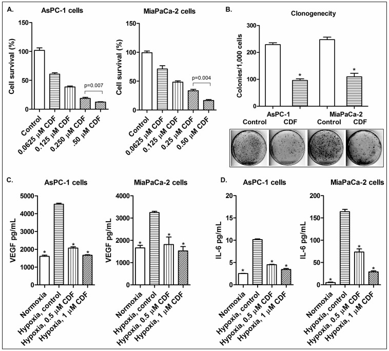
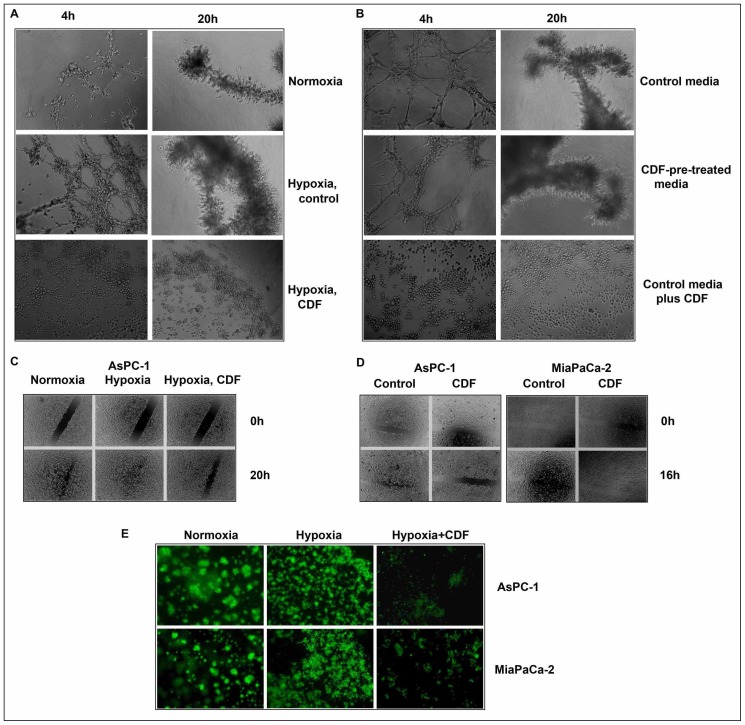
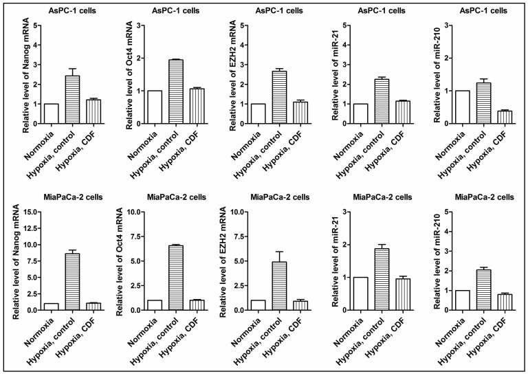
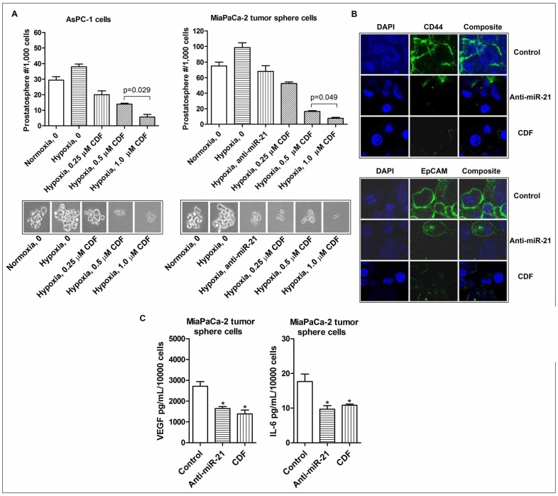
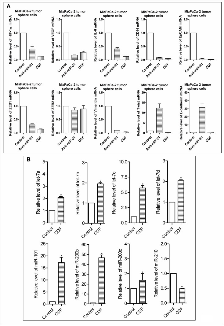
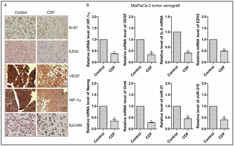
Hypoxia is known to play critical roles in cell survival, angiogenesis, tumor invasion, and metastasis. Hypoxia mediated over-expression of hypoxia-inducible factor (HIF) has been shown to be associated with therapeutic resistance, and contributes to poor prognosis of cancer patients. Emerging evidence suggest that hypoxia and HIF pathways contributes to the acquisition of epithelial-to-mesenchymal transition (EMT), maintenance of cancer stem cell (CSC) functions, and also maintains the vicious cycle of inflammation-all which lead to therapeutic resistance. However, the precise molecular mechanism(s) by which hypoxia/HIF drives these events are not fully understood. Here, we show, for the first time, that hypoxia leads to increased expression of VEGF, IL-6, and CSC signature genes Nanog, Oct4 and EZH2 consistent with increased cell migration/invasion and angiogenesis, and the formation of pancreatospheres, concomitant with increased expression of miR-21 and miR-210 in human pancreatic cancer (PC) cells. The treatment of PC cells with CDF, a novel synthetic compound inhibited the production of VEGF and IL-6, and down-regulated the expression of Nanog, Oct4, EZH2 mRNAs, as well as miR-21 and miR-210 under hypoxia. CDF also led to decreased cell migration/invasion, angiogenesis, and formation of pancreatospheres under hypoxia. Moreover, CDF decreased gene expression of miR-21, miR-210, IL-6, HIF-1α, VEGF, and CSC signatures in vivo in a mouse orthotopic model of human PC. Collectively, these results suggest that the anti-tumor activity of CDF is in part mediated through deregulation of tumor hypoxic pathways, and thus CDF could become a novel, and effective anti-tumor agent for PC therapy.


Conflict of interest statement

Competing Interests: Co-author Dr. Sarkar is a PLOS ONE Editorial Board member. This does not alter the authors' adherence to all the PLOS ONE policies on sharing data and materials.

Figures

Figure 1
Figure 1. Effect of CDF on cell growth, clonogenicity, and on the expression of VEGF and IL-6 in human PC cells under hypoxic conditions.
The panels A, B, C, and D represent cell growth, clonogenicity, VEGF, and IL-6, respectively. The conditioned media were collected from cells grown under normoxic and hypoxic conditions, as described under the methods section. The measurements of VEGF and IL-6 were conducted by ELISA. The bars in the charts indicate standard deviation (SD) of at least n = 3. * indicates p<0.05.
Figure 2
Figure 2. Effect of CDF on angiogenesis, cell migration, and invasion in PC cells under hypoxic conditions.
The panels A&B, C&D, and E represent angiogenesis, cell migration, and invasion, respectively. As described in the Methods section, angiogenesis in vitro was evaluated by the tube formation assay using endothelial cells; cell migration was evaluated by wound healing assay; invasion was evaluated by chamber invasion assay.
Figure 3
Figure 3. Effect of CDF on the expression of Nanog, Oct4, EZH2, miR-21, and miR-210 in PC cells under hypoxic conditions.
Real time RT-PCR was employed as described under the Methods section. The bars in the charts indicate SD of n = 3. * indicates p<0.05.
Figure 4
Figure 4. Effect of CDF and anti-miR-21 on the formation of pancreatospheres, the expression of CD44 and EpCAM, and the production of VEGF and IL-6 in the CSC-like sphere cells of PC cells under hypoxic conditions.
The panels A, B, and C represent the formation of pancreatospheres, the expression of CD44 and EpCAM, and the production of VEGF and IL-6, respectively. The sphere formation assay was conducted to examine the self-renewal capacity of CSCs in PC cells, as described under the Methods section. Confocal imaging microscopy (200× magnification) was conducted to measure CSC cell surface markers CD44 and EpCAM in the MiaPaCa-2 tumor sphere cells, as described under the Methods section. The bars in the charts indicate SD of n = 3. * indicates p<0.05.
Figure 5
Figure 5. Effect of CDF and/or anti-miR-21 on the mRNA and miRNA expressions in the CSC-like sphere cells of PC cells under hypoxic conditions.
The panels A and B represent the mRNAs and miRNAs, respectively. The sphere cells were transfected with anit-miR-21 by using the transfection reagent, following the manufacturer's manual. Real time RT-PCR was employed as described under the Methods section. The bars in the charts indicate SD of n = 3. * indicates p<0.05.
Figure 6
Figure 6. CDF decreased the expression of cell proliferation marker Ki-67, and other markers of tumor aggressiveness such as HIF-1α, VEGF, EZH2, EpCAM as well as the expression of HIF-1α, VEGF, IL-6, EZH2, Nanog, Oct4 mRNAs, miR-21, and miR-210 in pancreatic tumor tissues (orthotopic tumors induced by human MiaPaCa-2 cells).
The panels A and B represent immunohistochemistry assessed by immunohistochemistry (20× magnification), and mRNAs/miRNAs assessed by real time RT-PCR, respectively. The bars in the charts indicate SD of n = 3. * indicates p<0.05.

Similar articles

Cited by

References

    1. Siegel R, Naishadham D, Jemal A (2012) Cancer statistics, 2012. CA Cancer J Clin 62(1): 10–29. - PubMed
    1. Arslan AA, Helzlsouer KJ, Kooperberg C, Shu XO, Steplowski E, et al. (2010) Anthropometric measures, body mass index, and pancreatic cancer: a pooled analysis from the Pancreatic Cancer Cohort Consortium (PanScan). Arch Intern Med 170(9): 791–802. - PMC - PubMed
    1. Edwards BK, Brown ML, Wingo PA, Howe HL, Ward E, et al. (2005) Annual report to the nation on the status of cancer, 1975–2002, featuring population-based trends in cancer treatment. J Natl Cancer Inst 97(19): 1407–1427. - PubMed
    1. Jubb AM, Buffa FM, Harris AL (2010) Assessment of tumour hypoxia for prediction of response to therapy and cancer prognosis. J Cell Mol Med 14 1–2: 18–29. - PMC - PubMed
    1. Moulder JE, Rockwell S (1987) Tumor hypoxia: its impact on cancer therapy. Cancer Metastasis Rev 5 4: 313–341. - PubMed

Publication types

MeSH terms