

Mucin deficiency causes functional and structural changes of the ocular surface
- PMID: 23272068
- PMCID: PMC3525643
- DOI: 10.1371/journal.pone.0050704
Mucin deficiency causes functional and structural changes of the ocular surface
Abstract
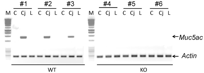
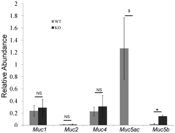
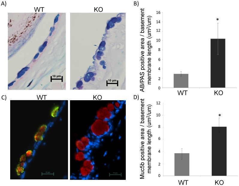
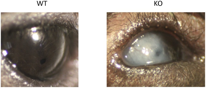
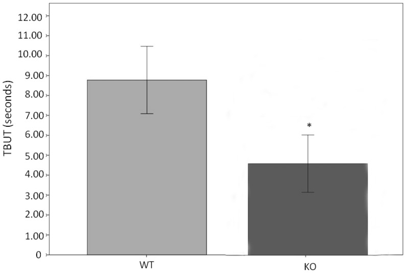
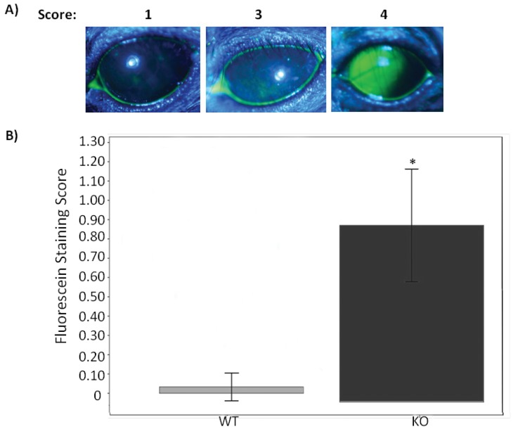
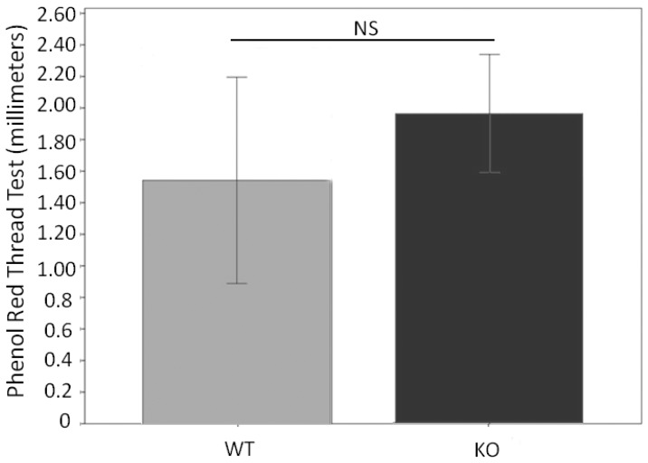
MUC5AC is the most abundant gel-forming mucin in the ocular system. However, the specific function is unknown. In the present study, a Muc5ac knockout (KO) mouse model was subject to various physiological measurements as compared to its wide-type (WT) control. Interestingly, when KO mice were compared to WT mice, the mean tear break up time (TBUT) values were significantly lower and corneal fluorescein staining scores were significantly higher. But the tear volume was not changed. Despite the lack of Muc5ac expression in the conjunctiva of KO mice, Muc5b expression was significantly increased in these mice. Corneal opacification, varying in location and severity, was found in a few KO mice but not in WT mice. The present results suggest a significant difference in the quality, but not the quantity, of tear fluid in the KO mice compared to WT mice. Dry eye disease is multifactorial and therefore further evaluation of the varying components of the tear film, lacrimal unit and corneal structure of these KO mice may help elucidate the role of mucins in dry eye disease. Because Muc5ac knockout mice have clinical features of dry eye, this mouse model will be extremely useful for further studies regarding the pathophysiology of the ocular surface in dry eye in humans.
Conflict of interest statement
Figures
Similar articles
-
The ocular surface phenotype of Muc5ac and Muc5b null mice.Invest Ophthalmol Vis Sci. 2014 Jan 15;55(1):291-300. doi: 10.1167/iovs.13-13194. Invest Ophthalmol Vis Sci. 2014. PMID: 24327612 Free PMC article.
-
Graft Versus Host Disease-Associated Dry Eye: Role of Ocular Surface Mucins and the Effect of Rebamipide, a Mucin Secretagogue.Invest Ophthalmol Vis Sci. 2019 Nov 1;60(14):4511-4519. doi: 10.1167/iovs.19-27843. Invest Ophthalmol Vis Sci. 2019. PMID: 31675422
-
Atopic ocular surface disease: implications on tear function and ocular surface mucins.Cornea. 2005 Nov;24(8 Suppl):S18-S23. doi: 10.1097/01.ico.0000178741.14212.53. Cornea. 2005. PMID: 16227818 Review.
-
Interleukin-1 receptor-1-deficient mice show attenuated production of ocular surface inflammatory cytokines in experimental dry eye.Cornea. 2008 Aug;27(7):811-7. doi: 10.1097/ICO.0b013e31816bf46c. Cornea. 2008. PMID: 18650668
-
Secreted Mucins on the Ocular Surface.Invest Ophthalmol Vis Sci. 2018 Nov 1;59(14):DES151-DES156. doi: 10.1167/iovs.17-23623. Invest Ophthalmol Vis Sci. 2018. PMID: 30481820 Review.
Cited by
-
The protective role of conjunctival goblet cell mucin sialylation.Nat Commun. 2023 Mar 17;14(1):1417. doi: 10.1038/s41467-023-37101-y. Nat Commun. 2023. PMID: 36932081 Free PMC article.
-
Characterisation of Gel-Forming Mucins Produced In Vivo and In Ex Vivo Conjunctival Explant Cultures.Int J Mol Sci. 2021 Sep 29;22(19):10528. doi: 10.3390/ijms221910528. Int J Mol Sci. 2021. PMID: 34638869 Free PMC article.
-
Mucus models to evaluate the diffusion of drugs and particles.Adv Drug Deliv Rev. 2018 Jan 15;124:34-49. doi: 10.1016/j.addr.2017.11.001. Epub 2017 Nov 5. Adv Drug Deliv Rev. 2018. PMID: 29117512 Free PMC article. Review.
-
Dry Eye Etiology: Focus on Friction.Klin Monbl Augenheilkd. 2020 Oct;237(10):1235-1236. doi: 10.1055/a-0898-3857. Epub 2019 Jul 30. Klin Monbl Augenheilkd. 2020. PMID: 31362317 Free PMC article. No abstract available.
-
Protective Role of Surfactant Protein D in Ocular Staphylococcus aureus Infection.PLoS One. 2015 Sep 23;10(9):e0138597. doi: 10.1371/journal.pone.0138597. eCollection 2015. PLoS One. 2015. PMID: 26398197 Free PMC article.
References
-
- Gum JR Jr (1995) Human mucin glycoproteins: varied structures predict diverse properties and specific functions. Biochemical Society Transactions 23: 795–799. - PubMed
-
- Moniaux N, Escande F, Porchet N, Aubert JP, Batra SK (2001) Structural organization and classification of the human mucin genes. Front Biosci 6: D1192–1206. - PubMed
-
- Desseyn JL, Aubert JP, Porchet N, Laine A (2000) Evolution of the large secreted gel-forming mucins. Molecular Biology and Evolution 17: 1175–1184. - PubMed
-
- Offner GD, Troxler RF (2000) Heterogeneity of high-molecular-weight human salivary mucins. Adv Dent Res 14: 69–75. - PubMed
-
- Katsumi A, Tuley EA, Bodó I, Sadler JE (2000) Localization of disulfide bonds in the cystine knot domain of human von Willebrand factor. Journal of Biological Chemistry 275: 25585–25594. - PubMed
Publication types
MeSH terms
Substances
Grants and funding
LinkOut - more resources
Full Text Sources
Other Literature Sources
Medical
Molecular Biology Databases
Research Materials
