. 2012;7(12):e51524.
doi: 10.1371/journal.pone.0051524. Epub 2012 Dec 13.

δ-opioid receptor activation and microRNA expression of the rat cortex in hypoxia

Affiliations

δ-opioid receptor activation and microRNA expression of the rat cortex in hypoxia

Yilin Yang et al. PLoS One. 2012.

Abstract

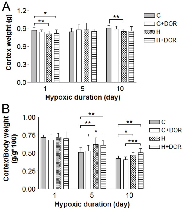
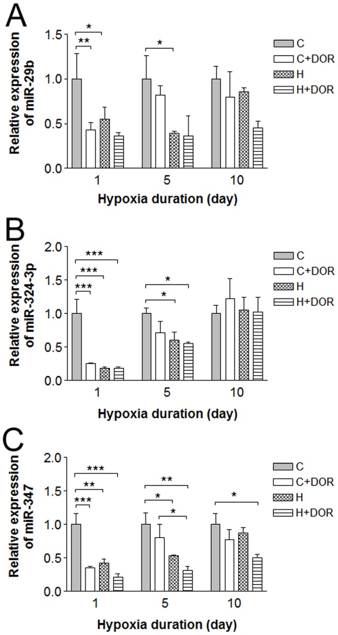
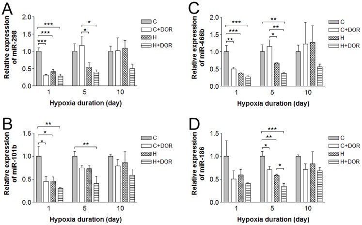
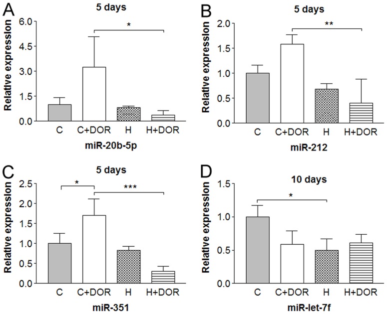
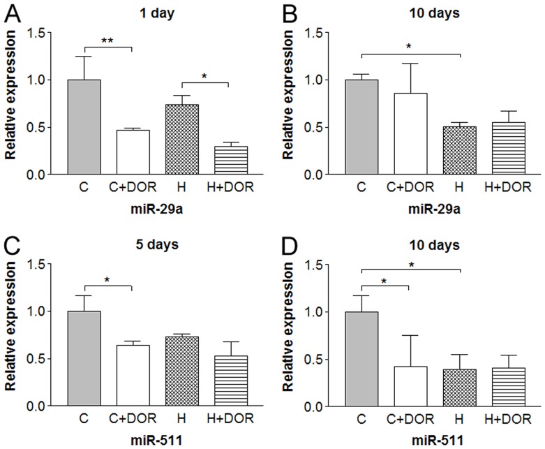
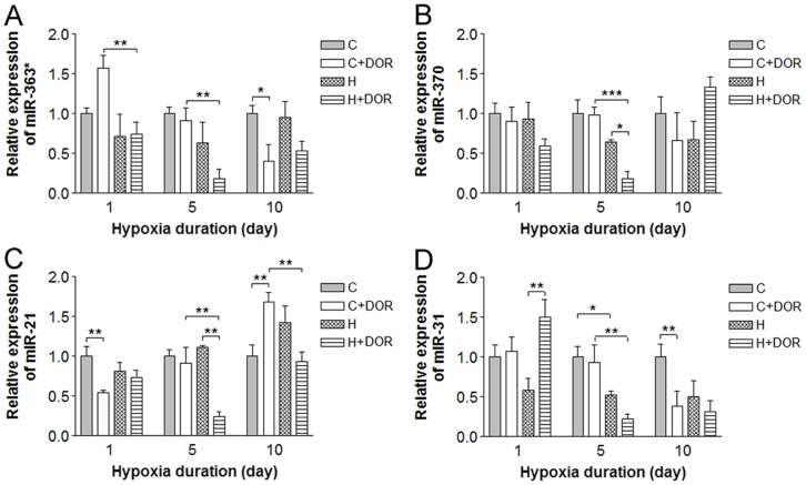
Prolonged hypoxic/ischemic stress may cause cortical injury and clinically manifest as a neurological disability. Activation of the δ-opioid receptor (DOR) may induce cortical protection against hypoxic/ischemic insults. However, the mechanisms underlying DOR protection are not clearly understood. We have recently found that DOR activation modulates the expression of microRNAs (miRNAs) in the kidney exposed to hypoxia, suggesting that DOR protection may involve a miRNA mechanism. To determine if the miRNAs expressed in the cortex mediated DOR neuroprotection, we examined 19 miRNAs that were previously identified as hypoxia- and DOR-regulated miRNAs in the kidney, in the rat cortex treated with UFP-512, a potent and specific DOR agonist under hypoxic condition. Of the 19 miRNAs tested, 17 were significantly altered by hypoxia and/or DOR activation with the direction and amplitude varying depending on hypoxic duration and times of DOR treatment. Expression of several miRNAs such as miR-29b, -101b, -298, 324-3p, -347 and 466b was significantly depressed after 24 hours of hypoxia. Similar changes were seen in normoxic condition 24 hours after DOR activation with one-time treatment of UFP-512. In contrast, some miRNAs were more tolerant to hypoxic stress and showed significant reduction only with 5-day (e.g., miR-31 and -186) or 10-day (e.g., miR-29a, let-7f and -511) exposures. In addition, these miRNAs had differential responses to DOR activation. Other miRNAs like miRs-363* and -370 responded only to the combined exposure to hypoxia and DOR treatment, with a notable reduction of >70% in the 5-day group. These data suggest that cortical miRNAs are highly yet differentially sensitive to hypoxia. DOR activation can modify, enhance or resolve the changes in miRNAs that target HIF, ion transport, axonal guidance, free radical signaling, apoptosis and many other functions.


Conflict of interest statement

Competing Interests: The authors have declared that no competing interests exist.

Figures

Figure 1
Figure 1. Effect of hypoxia and DOR activation on the cortex and body weight.
A, Cortex weight (n = 9). B, Ratio of cortex to body weight for each study group (n = 6–9). C: control; C+DOR: control+DOR agonist UFP-512; H: hypoxia; H+DOR: hypoxia+ DOR agonist UFP-512. *p<0.05, **p<0.01, ***p<0.001. Note that small, but significant, drops in cortex weight were found in both 1-day and 10-day hypoxic animals, and the cortex/body weight ratio increased with 5–10 day hypoxia. Also note that UFP-512 did not have a detectable affect on cortex weights, but preserved more of the cortex/body weight ratio.
Figure 2
Figure 2. Relative miRNA expression levels of miR-29b, -324-3p and -347 in the cortex following 1, 5 or 10 days of hypoxia or UFP-512 treatment.
C: control; C+DOR: control+DOR agonist UFP-512; H: hypoxia; H+DOR: hypoxia+ DOR agonist UFP-512. *p<0.05, **p<0.01, ***p<0.001. Note that miRNA expression levels of miR-29b, -324-3p and -347 were suppressed in earlier, but not later days (10-day) of hypoxia. UFP-512 suppressed these miRNAs’ expression in a similar way. These results suggest these miRNAs respond to hypoxia and DOR activation in the early period, but not in the extended duration.
Figure 3
Figure 3. Relative miRNA expression levels of miR-298, -466b, -101b and -186 in the rat cortex following 1, 5 or 10 days of hypoxia or UFP-512 treatment.
C: control; C+DOR: control+DOR agonist UFP-512; H: hypoxia; H+DOR: hypoxia+ DOR agonist UFP-512. *p<0.05, **p<0.01, ***p<0.001. Note that miRs-298, 446b and 101b are all significantly downregulated in response to hypoxia or UFP-512 alone within the first 24 hours of treatment. Hypoxia reduced the level of miR-186 after 1-day exposure and this change persisted and reached significance after 5 days of exposure to hypoxia.
Figure 4
Figure 4. Relative miRNA expression levels of miR-20b-5p, -212, -351 and miR-let-7f in the rat cortex following 5 or 10 days of hypoxia.
C: control; C+DOR: control+DOR agonist UFP-512; H: hypoxia; H+DOR: hypoxia+ DOR agonist UFP-512. *p<0.05, **p<0.01, ***p<0.001. Note that the expression of miRs-20b-5p, -212 and -351 was not significantly affected by 5 days of hypoxia alone, but significantly suppressed in the co-presence of hypoxia and UFP-512. The expression of miR-let-7f was significantly reduced by 10-day exposure to hypoxic conditions, but was not affected by UFP-512.
Figure 5
Figure 5. Relative miRNA expression levels of miR-29a and miR-511 in the rat cortex following 1, 5 or 10 days of hypoxia with or without UFP-512 treatment.
C: control; C+DOR: control+DOR agonist UFP-512; H: hypoxia; H+DOR: hypoxia+ DOR agonist UFP-512. *p<0.05, **p<0.01. Note that miRNA expression levels of miR-29a and miR-511 were altered at discrete time points by hypoxia and/or UFP-512.
Figure 6
Figure 6. Relative miRNA expression levels of miR-363*, -370, -21 and miR-31 in the rat cortex following 1, 5 or 10 days of hypoxia with or without UFP-512 treatment.
C: control; C+DOR: control+DOR agonist UFP-512; H: hypoxia; H+DOR: hypoxia+ DOR agonist UFP-512. *p<0.05, **p<0.01, ***p<0.001. Note that combination of hypoxia and UFP-512 treatment significantly altered the expression levels of miR-363*, -370, -21 and miR-31 in the cortex following 10-day treatment.

References

    1. Zhang J, Haddad GG, Xia Y (2000) delta-, but not mu- and kappa-, opioid receptor activation protects neocortical neurons from glutamate-induced excitotoxic injury. Brain Res 885: 143–153. - PubMed
    1. Zhang J, Gibney GT, Zhao P, Xia Y (2002) Neuroprotective role of delta-opioid receptors in cortical neurons. Am J Physiol Cell Physiol 282: C1225–1234. - PubMed
    1. Ma MC, Qian H, Ghassemi F, Zhao P, Xia Y (2005) Oxygen-sensitive {delta}-opioid receptor-regulated survival and death signals: novel insights into neuronal preconditioning and protection. J Biol Chem 280: 16208–16218. - PubMed
    1. Zhang J, Qian H, Zhao P, Hong SS, Xia Y (2006) Rapid hypoxia preconditioning protects cortical neurons from glutamate toxicity through delta-opioid receptor. Stroke 37: 1094–1099. - PubMed
    1. Chao D, Donnelly DF, Feng Y, Bazzy-Asaad A, Xia Y (2007) Cortical delta-opioid receptors potentiate K+ homeostasis during anoxia and oxygen-glucose deprivation. J Cereb Blood Flow Metab 27: 356–368. - PubMed

Publication types

Substances