

Effects of exercise on AMPK signaling and downstream components to PI3K in rat with type 2 diabetes
- PMID: 23272147
- PMCID: PMC3521695
- DOI: 10.1371/journal.pone.0051709
Effects of exercise on AMPK signaling and downstream components to PI3K in rat with type 2 diabetes
Abstract
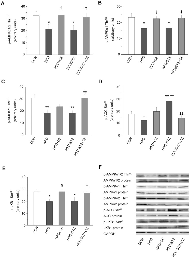
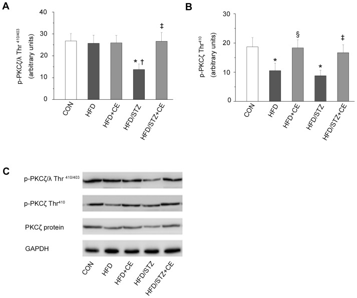
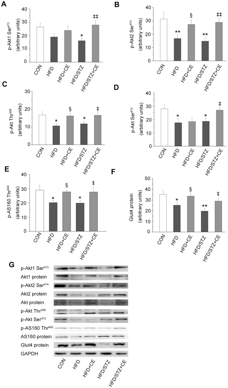
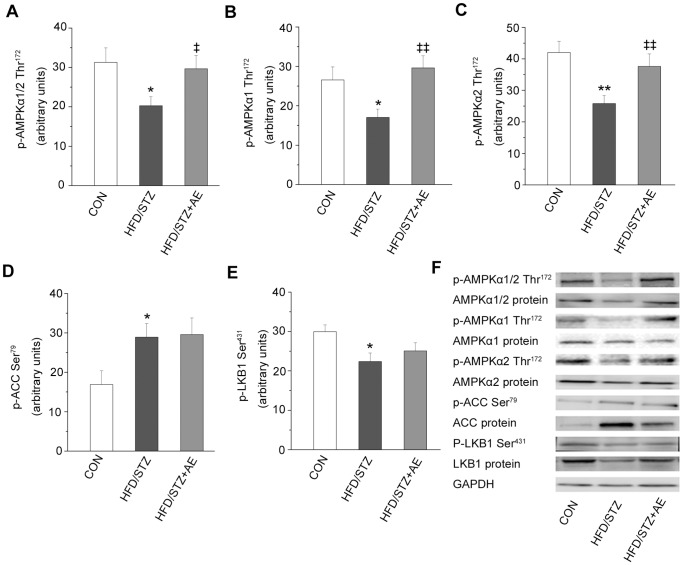
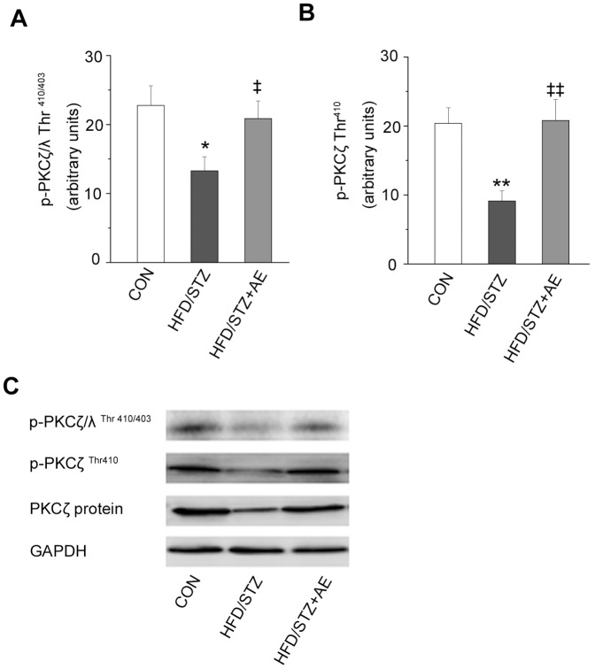
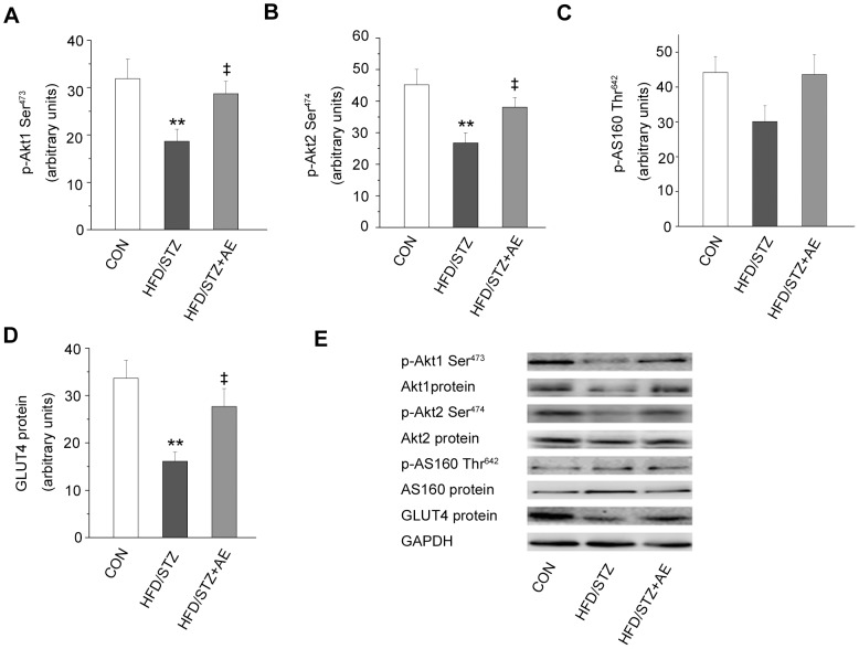
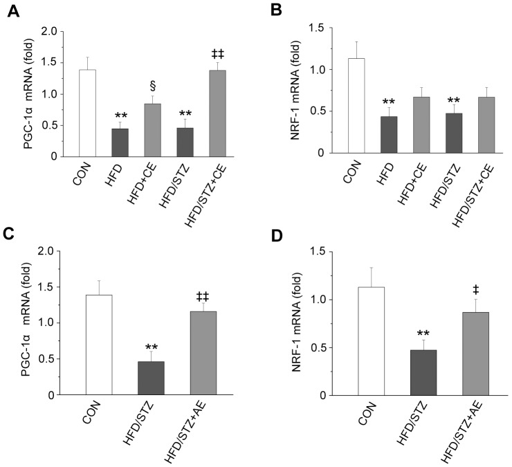
Exercise can increase skeletal muscle sensitivity to insulin, improve insulin resistance and regulate glucose homeostasis in rat models of type 2 diabetes. However, the potential mechanism remains poorly understood. In this study, we established a male Sprague-Dawley rat model of type 2 diabetes, with insulin resistance and β cell dysfunction, which was induced by a high-fat diet and low-dose streptozotocin to replicate the pathogenesis and metabolic characteristics of type 2 diabetes in humans. We also investigated the possible mechanism by which chronic and acute exercise improves metabolism, and the phosphorylation and expression of components of AMP-activated protein kinase (AMPK) and downstream components of phosphatidylinositol 3-kinase (PI3K) signaling pathways in the soleus. As a result, blood glucose, triglyceride, total cholesterol, and free fatty acid were significantly increased, whereas insulin level progressively declined in diabetic rats. Interestingly, chronic and acute exercise reduced blood glucose, increased phosphorylation and expression of AMPKα1/2 and the isoforms AMPKα1 and AMPKα2, and decreased phosphorylation and expression of AMPK substrate, acetyl CoA carboxylase (ACC). Chronic exercise upregulated phosphorylation and expression of AMPK upstream kinase, LKB1. But acute exercise only increased LKB1 expression. In particular, exercise reversed the changes in protein kinase C (PKC)ζ/λ phosphorylation, and PKCζ phosphorylation and expression. Additionally, exercise also increased protein kinase B (PKB)/Akt1, Akt2 and GLUT4 expression, but AS160 protein expression was unchanged. Chronic exercise elevated Akt (Thr(308)) and (Ser(473)) and AS160 phosphorylation. Finally, we found that exercise increased peroxisome proliferator-activated receptor-γ coactivator 1 (PGC1) mRNA expression in the soleus of diabetic rats. These results indicate that both chronic and acute exercise influence the phosphorylation and expression of components of the AMPK and downstream to PIK3 (aPKC, Akt), and improve GLUT4 trafficking in skeletal muscle. These data help explain the mechanism how exercise regulates glucose homeostasis in diabetic rats.
Conflict of interest statement
Figures
References
-
- Højlund K, Mogensen M, Sahlin K, Beck-Nielsen H (2008) Mitochondrial dysfunction in type 2 diabetes and obesity. Endocrinol Metab Clin North Am 37: 713–731, x. - PubMed
-
- Kim CH, Youn JH, Park JY, Hong SK, Park KS, et al. (2000) Effects of high-fat diet and exercise training on intracellular glucose metabolism in rats. Am J Physiol Endocrinol Metab 278: E977–984. - PubMed
-
- Kraegen EW, Storlien LH, Jenkins AB, James DE (1989) Chronic exercise compensates for insulin resistance induced by a high-fat diet in rats. Am J Physiol 256: E242–249. - PubMed
-
- Lessard SJ, Rivas DA, Chen ZP, Bonen A, Febbraio MA, et al. (2007) Tissue-specific effects of rosiglitazone and exercise in the treatment of lipid-induced insulin resistance. Diabetes 56: 1856–1864. - PubMed
-
- Srinivasan K, Viswanad B, Asrat L, Kaul CL, Ramarao P (2005) Combination of high-fat diet-fed and low-dose streptozotocin-treated rat: a model for type 2 diabetes and pharmacological screening. Pharmacol Res 52: 313–320. - PubMed
Publication types
MeSH terms
Substances
LinkOut - more resources
Full Text Sources
Medical
Molecular Biology Databases
Research Materials
Miscellaneous
