. 2012;7(12):e51829.
doi: 10.1371/journal.pone.0051829. Epub 2012 Dec 13.

The role of human Dicer-dsRBD in processing small regulatory RNAs

Affiliations

The role of human Dicer-dsRBD in processing small regulatory RNAs

Christopher Wostenberg et al. PLoS One. 2012.

Abstract

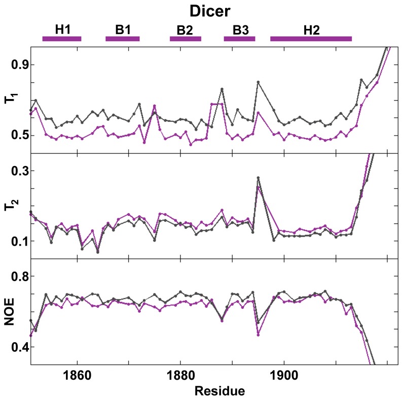
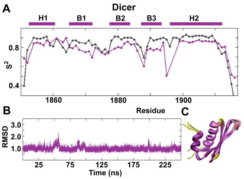
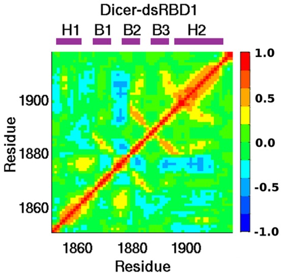
One of the most exciting recent developments in RNA biology has been the discovery of small non-coding RNAs that affect gene expression through the RNA interference (RNAi) mechanism. Two major classes of RNAs involved in RNAi are small interfering RNA (siRNA) and microRNA (miRNA). Dicer, an RNase III enzyme, plays a central role in the RNAi pathway by cleaving precursors of both of these classes of RNAs to form mature siRNAs and miRNAs, which are then loaded into the RNA-induced silencing complex (RISC). miRNA and siRNA precursors are quite structurally distinct; miRNA precursors are short, imperfect hairpins while siRNA precursors are long, perfect duplexes. Nonetheless, Dicer is able to process both. Dicer, like the majority of RNase III enzymes, contains a dsRNA binding domain (dsRBD), but the data are sparse on the exact role this domain plays in the mechanism of Dicer binding and cleavage. To further explore the role of human Dicer-dsRBD in the RNAi pathway, we determined its binding affinity to various RNAs modeling both miRNA and siRNA precursors. Our study shows that Dicer-dsRBD is an avid binder of dsRNA, but its binding is only minimally influenced by a single-stranded - double-stranded junction caused by large terminal loops observed in miRNA precursors. Thus, the Dicer-dsRBD contributes directly to substrate binding but not to the mechanism of differentiating between pre-miRNA and pre-siRNA. In addition, NMR spin relaxation and MD simulations provide an overview of the role that dynamics contribute to the binding mechanism. We compare this current study with our previous studies of the dsRBDs from Drosha and DGCR8 to give a dynamic profile of dsRBDs in their apo-state and a mechanistic view of dsRNA binding by dsRBDs in general.


Conflict of interest statement

Competing Interests: The authors have declared that no competing interests exist.

Figures

Figure 1
Figure 1. Schematic representation and structure of Dicer-dsRBD.
(A) Schematic representation of the primary sequence of Dicer with approximate location of domains above the cartoon. The primary sequence of human Dicer-dsRBD (residues 1850 to 1922) utilized in this study is shown in the box below the schematic, with the approximate location of secondary elements shown above the sequence(H represents an α-helix, B a β-sheet, and L a loop/turn). (B) Cryo-EM reconstruction is shown with the surface colored gray and the individual domains colored the same as in the above schematic. The tertiary structure of the mouse Dicer-dsRBD (pdb 3C4B, residues 1833 to 1900), which is 100% identical to the human sequence (residues 1849 to 1916), is depicted in the box to the right of the cryo-EM reconstruction.
Figure 2
Figure 2. EMSA of pre-mir-16-1 and ds44 binding by Dicer-dsRBD.
EMSA of Dicer-dsRBD binding (A) pre-mir-16-1 with a Kd = 2.2±0.1 µM and (B) ds44 with a Kd = 2.4±0.1 µM. The predicted secondary structures of the RNAs are shown above the representative gels, which were run with varying Dicer-dsRBD concentration (0.25–50.12 µM) binding to 0.125 nM RNA. Fraction bound, from the EMSA data, versus Dicer-dsRBD concentration was fitted using a generalized Hill model (gray line). Error bars in the plots of fraction bound as a function of total Dicer-dsRBD concentration represent the standard deviation from duplicate measurements.
Figure 3
Figure 3. Sedimentation velocity analysis of Dicer-dsRBD binding to ds16.
Plots of normalized g(s*) distributions for 2.0 µM ds16 alone (black) and 2.0 µM ds16 plus 4.3 µM (red), 8.5 µM (blue), 21.4 µM (green), and 42.7 µM (lavender) Dicer-dsRBD. The distributions are normalized by area. The shift in the peak position from 2.3 S for the RNA alone to 3.3 S for the complex corresponds to a one-to-one binding stoichiometry. The data were globally analyzed using a one-to-one binding model to yield a best fit Kd = 5.4±0.7 µM, which agrees well with the EMSA data.
Figure 4
Figure 4. EMSA of ds16-tetra-stable and ds16-octa-U binding by Dicer-dsRBD.
EMSA of Dicer-dsRBD binding (A) ds16-tetra-stable with a Kd = 9.1±0.1 µM and (B) ds16-octa-U with a Kd = 4.7±0.1 µM. The predicted secondary structures of the RNAs are shown above the representative gels, which were run with various Dicer-dsRBD concentration (0.25–50.12 µM) binding to 0.125 nM RNA. Fraction bound, from the EMSA data, versus Dicer-dsRBD concentration was fitted using a generalized Hill cooperative model (gray line). Error bars in the plots of fraction bound as a function of total Dicer-dsRBD concentration represent the standard deviation from duplicate measurements.
Figure 5
Figure 5. NMR titration of Dicer-dsRBD with ds33.
Representative 15N-HSQC spectra of Dicer-dsRBD collected in the unbound state and in the presence of ds33. (A) Reference spectrum of apo-Dicer-dsRBD. (B) Ratio of individual peak intensities in the presence of 0.02∶1 mole ratio ds33:Dicer-dsRBD to those recorded under identical conditions in the apo-state spectrum displayed in (A). (C) Representative spectra from the ds33 titration showing the data points with mole ratios of 0.02∶1, 0.20∶1, and 2.0∶1 ds33:Dicer-dsRBD as labeled. All spectra were collected at 25°C in the presence of 100 mM KCl on a spectrometer operating at 600 MHz field strength.
Figure 6
Figure 6. 15N spin relaxation experiments of Dicer-dsRBD.
15N spin relaxation data for Dicer-dsRBD collected at 500 MHz (purple) and 600 MHz (gray) shows that the most dynamic regions of Dicer-dsRBD on the picosecond to nanosecond timescale are the loops, most notably loop 4. The secondary elements are represented as purple bars above the plot.
Figure 7
Figure 7. Order parameters and RMSD of Dicer-dsRBD along with PDB bundle.
(A) Order parameters (S2) plotted for Dicer-dsRBD show that the most flexible regions in the protein are loops 3 and 4. Experimental data (purple) is plotted with MD predicted order parameters (gray). The secondary elements are represented as purple bars above the plot. (B) The overall stability of Dicer-dsRBD during the 250 ns MD simulation is demonstrated by the low average RMSD (<1.0 Å). (C) MD-derived ribbon bundle for Dicer-dsRBD also shows the stability of the construct. Increased flexibility, derived from the experimental order parameters, is depicted colorimetrically on the ribbon bundle as passage from purple (high order parameter) to yellow (low order parameter). The bundle was created by taking structures from the simulation every 50 ns and superimposing them to remove translational and rotational motion.
Figure 8
Figure 8. Cα correlations of Dicer-dsRBD.
The Cα correlation matrix of Dicer-dsRBD reveals backbone motions. The color bar on the right shows the scale indicating strong positive correlation (red), strong negative correlation (blue), and non-correlated (green) motions. Labels above the panel indicate the location of secondary structural elements within the sequence.

References

    1. Kim VN, Han J, Siomi MC (2009) Biogenesis of small RNAs in animals. Nat Rev Mol Cell Biol 10: 126–139. - PubMed
    1. Jinek M, Doudna JA (2009) A three-dimensional view of the molecular machinery of RNA interference. Nature 457: 405–412. - PubMed
    1. van Rij RP, Berezikov E (2009) Small RNAs and the control of transposons and viruses in Drosophila. Trends Microbiol 17: 163–171. - PubMed
    1. Song L, Gao S, Jiang W, Chen S, Liu Y, et al. (2011) Silencing suppressors: viral weapons for countering host cell defenses. Protein Cell 2: 273–281. - PMC - PubMed
    1. Nimmo RA, Slack FJ (2009) An elegant miRror: microRNAs in stem cells, developmental timing and cancer. Chromosoma 118: 405–418. - PMC - PubMed

Publication types

MeSH terms