. 2012;7(12):e51930.
doi: 10.1371/journal.pone.0051930. Epub 2012 Dec 13.

Differential requirement for protein synthesis in presynaptic unmuting and muting in hippocampal glutamate terminals

Affiliations

Differential requirement for protein synthesis in presynaptic unmuting and muting in hippocampal glutamate terminals

Devon C Crawford et al. PLoS One. 2012.

Abstract

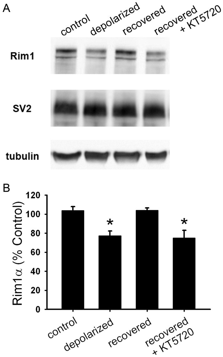
Synaptic function and plasticity are crucial for information processing within the nervous system. In glutamatergic hippocampal neurons, presynaptic function is silenced, or muted, after strong or prolonged depolarization. This muting is neuroprotective, but the underlying mechanisms responsible for muting and its reversal, unmuting, remain to be clarified. Using cultured rat hippocampal neurons, we found that muting induction did not require protein synthesis; however, slow forms of unmuting that depend on protein kinase A (PKA), including reversal of depolarization-induced muting and forskolin-induced unmuting of basally mute synapses, required protein synthesis. In contrast, fast unmuting of basally mute synapses by phorbol esters was protein synthesis-independent. Further studies of recovery from depolarization-induced muting revealed that protein levels of Rim1 and Munc13-1, which mediate vesicle priming, correlated with the functional status of presynaptic terminals. Additionally, this form of unmuting was prevented by both transcription and translation inhibitors, so proteins are likely synthesized de novo after removal of depolarization. Phosphorylated cyclic adenosine monophosphate response element-binding protein (pCREB), a nuclear transcription factor, was elevated after recovery from depolarization-induced muting, consistent with a model in which PKA-dependent mechanisms, possibly including pCREB-activated transcription, mediate slow unmuting. In summary, we found that protein synthesis was required for slower, PKA-dependent unmuting of presynaptic terminals, but it was not required for muting or a fast form of unmuting. These results clarify some of the molecular mechanisms responsible for synaptic plasticity in hippocampal neurons and emphasize the multiple mechanisms by which presynaptic function is modulated.


Conflict of interest statement

Competing Interests: The authors have declared that no competing interests exist.

Figures

Figure 1
Figure 1. Protein synthesis is not required for depolarization-induced muting. A.
Representative autaptic EPSCs from hippocampal neurons after 16 h 30 mM NaCl (control) or 30 mM KCl (depolarized) with or without 1 µg/ml cycloheximide 30 min pretreatment and co-incubation (cyh). B. Summary of EPSC amplitudes from neurons treated as in panel A (n = 10 neurons). *p<0.05, Bonferroni corrected Student’s unpaired t test.
Figure 2
Figure 2. Protein synthesis is required for functional recovery from depolarization-induced muting.
A. Representative autaptic EPSCs from hippocampal neurons after 16 h 30 mM NaCl (control), 16 h 30 mM KCl (depolarized), or 16 h 30 mM KCl followed by 3 h recovery in fresh medium with (recovered+cyh) or without (recovered) 1–5 µg/ml cycloheximide. Cycloheximide was applied 0.5–2 h prior to and during recovery. B. Summary of EPSC amplitudes from neurons treated as in panel A (n = 14 neurons). C. Representative autaptic EPSCs from hippocampal neurons after 16 h 30 mM NaCl (control), 16 h 30 mM KCl followed by 3 h recovery in fresh medium with 5 µg/ml cycloheximide (recovered+cyh), or 16 h 30 mM KCl followed by 3 h recovery in fresh medium with 5 µg/ml cycloheximide followed by an additional 3 h recovery in fresh medium without cycloheximide (recovered+cyh+recovery). D. Summary of autaptic EPSC amplitudes from neurons treated as in panel C (n = 25 neurons). *p<0.05, Bonferroni corrected Student’s unpaired t test.
Figure 3
Figure 3. Synthesis is required for protein recovery from depolarization-induced muting. A.
Western blot analysis of whole-cell lysates from hippocampal mass cultures treated with 16 h 30 mM NaCl (control), 16 h 30 mM KCl (depolarized), or 16 h 30 mM KCl followed by 3 h recovery in fresh medium with (recovered+cyh) or without (recovered) 1 µg/ml cycloheximide. Cycloheximide was applied 0.5 h prior to and during recovery. B. Summary of Rim1 levels from Western blots as shown in A (n = 3). Rim1 protein levels for each condition were normalized to SV2 and control treatment. C. Summary of Munc13–1 levels from Western blots as shown in A (n = 3). Munc13–1 protein levels for each condition were normalized to SV2 and control treatment. D. Representative images of Rim1 immunostaining in mass cultures after treatments as described in panel A. Scale bar represents 2 µm. E. Quantification of Rim1 immunostaining at vGluT-1-positive synapses (not shown; n = 15 fields). Integrated intensity values were normalized to the average control value for a given experiment. *p<0.05, Newman-Keuls post hoc test vs. control after one-way ANOVA.
Figure 4
Figure 4. PKA signaling is required for recovery of Rim1 levels after depolarization-induced muting. A.
Western blot analysis of whole-cell lysates from hippocampal mass cultures treated with 16 h 30 mM NaCl (control), 16 h 30 mM KCl (depolarized), or 16 h 30 mM KCl followed by 3 h recovery in fresh medium with (recovered+KT5720) or without (recovered) 2 µM KT5720. KT5720 was applied 0.5 h prior to and during recovery. B. Summary of Rim1 levels from Western blots as shown in A (n = 3). Rim1 protein levels for each condition were normalized to SV2 and control treatment. *p<0.05, Newman-Keuls post hoc test vs. control after one-way ANOVA.
Figure 5
Figure 5. Transcription is required for functional recovery from depolarization-induced muting. A.
Representative autaptic EPSCs from hippocampal neurons after 16 h 30 mM NaCl (control), 16 h 30 mM KCl (depolarized), or 16 h 30 mM KCl followed by 3 h recovery in fresh medium with (recovered+act D) or without (recovered) 200 ng/ml actinomycin D. Actinomycin D was applied 0.5 h prior to and during recovery. B. Summary of EPSC amplitudes from neurons treated as in panel A (n = 32 neurons). C. Quantification of Rim1 immunostaining at vGluT-1-positive synapses (not shown; n = 15 fields). Integrated intensity values were normalized to the average control value for a given experiment. *p<0.05, Bonferroni corrected Student’s one-tailed unpaired t test.
Figure 6
Figure 6. Nuclear phosphorylated CREB (pCREB) levels remain elevated after recovery from depolarization-induced muting. A.
Merged image of pCREB (red), GABA (blue), and MAP2 (green) immunofluorescence in a mass hippocampal culture. B. pCREB immunostaining after the following treatments: 30 min DMSO (control), 30 min 50 µM forskolin (forskolin 30 min), 30 min 30 mM KCl (depolarized 30 min), 16 h 30 mM KCl (depolarized O/N), or 16 h 30 mM KCl followed by 3 h in fresh medium (recovered). C. Quantification of pCREB intensity in GABA-negative nuclei after treatments as described in panel A (n = 327–586 neurons). Intensity values were normalized to the average control value for each experiment. *p<0.05, Student’s unpaired t test vs. control after Bonferroni correction. D. Quantification of pCREB intensity in GABA-negative nuclei after 16 h 30 mM NaCl (control), 16 h 30 mM KCl (depolarized O/N), or 16 h 30 mM KCl followed by 3 h recovery in fresh medium with (recovered+KT) or without (recovered) 2 µM KT5720 (n = 45–145 neurons). KT5720 was applied 0.5 h prior to and during recovery. Intensity values were normalized to the average control value for each experiment. *p<0.05, Bonferroni corrected Student’s unpaired t test.
Figure 7
Figure 7. Unmuting of basally mute synapses by forskolin, but not PDBu, requires protein synthesis.
A. Representative FM1-43FX (green) and vGluT-1 (red) merged images after 4 h DMSO (control), 4 hr DMSO plus 1 µg/ml cycloheximide (cyh), 4 h 50 µM forskolin (FSK 4 h), 4 h 50 µM forskolin plus 1 µg/ml cycloheximide (FSK 4 h+cyh), 2 min 1 µM PDBu (PDBu 2 min), or 2 min 1 µM PDBu plus 1 µg/ml cycloheximide (PDBu 2 min+cyh) treatment. Cycloheximide was applied 0.5 h prior to and during treatments. Scale bar represents 5 µm. B. Quantification of the percentage of vGluT-1-defined synapses labeled with FM1-43FX (active synapses) after treatments described in panel A (n = 20 fields). *p<0.05, Bonferroni corrected Student’s unpaired t test vs. control.

References

    1. Crawford DC, Mennerick S (2012) Presynaptically Silent Synapses: Dormancy and Awakening of Presynaptic Vesicle Release. Neuroscientist 18: 216–223. - PMC - PubMed
    1. Ninan I, Arancio O (2004) Presynaptic CaMKII is necessary for synaptic plasticity in cultured hippocampal neurons. Neuron 42: 129–141. - PubMed
    1. Ting JT, Kelley BG, Lambert TJ, Cook DG, Sullivan JM (2007) Amyloid precursor protein overexpression depresses excitatory transmission through both presynaptic and postsynaptic mechanisms. Proc Natl Acad Sci U S A 104: 353–358. - PMC - PubMed
    1. Moulder KL, Meeks JP, Shute AA, Hamilton CK, de Erausquin G, et al. (2004) Plastic elimination of functional glutamate release sites by depolarization. Neuron 42: 423–435. - PubMed
    1. Kohara K, Ogura A, Akagawa K, Yamaguchi K (2001) Increase in number of functional release sites by cyclic AMP-dependent protein kinase in cultured neurons isolated from hippocampal dentate gyrus. Neurosci Res 41: 79–88. - PubMed

Publication types