

Differences between the glycosylation patterns of haptoglobin isolated from skin scales and plasma of psoriatic patients
- PMID: 23272204
- PMCID: PMC3525549
- DOI: 10.1371/journal.pone.0052040
Differences between the glycosylation patterns of haptoglobin isolated from skin scales and plasma of psoriatic patients
Abstract
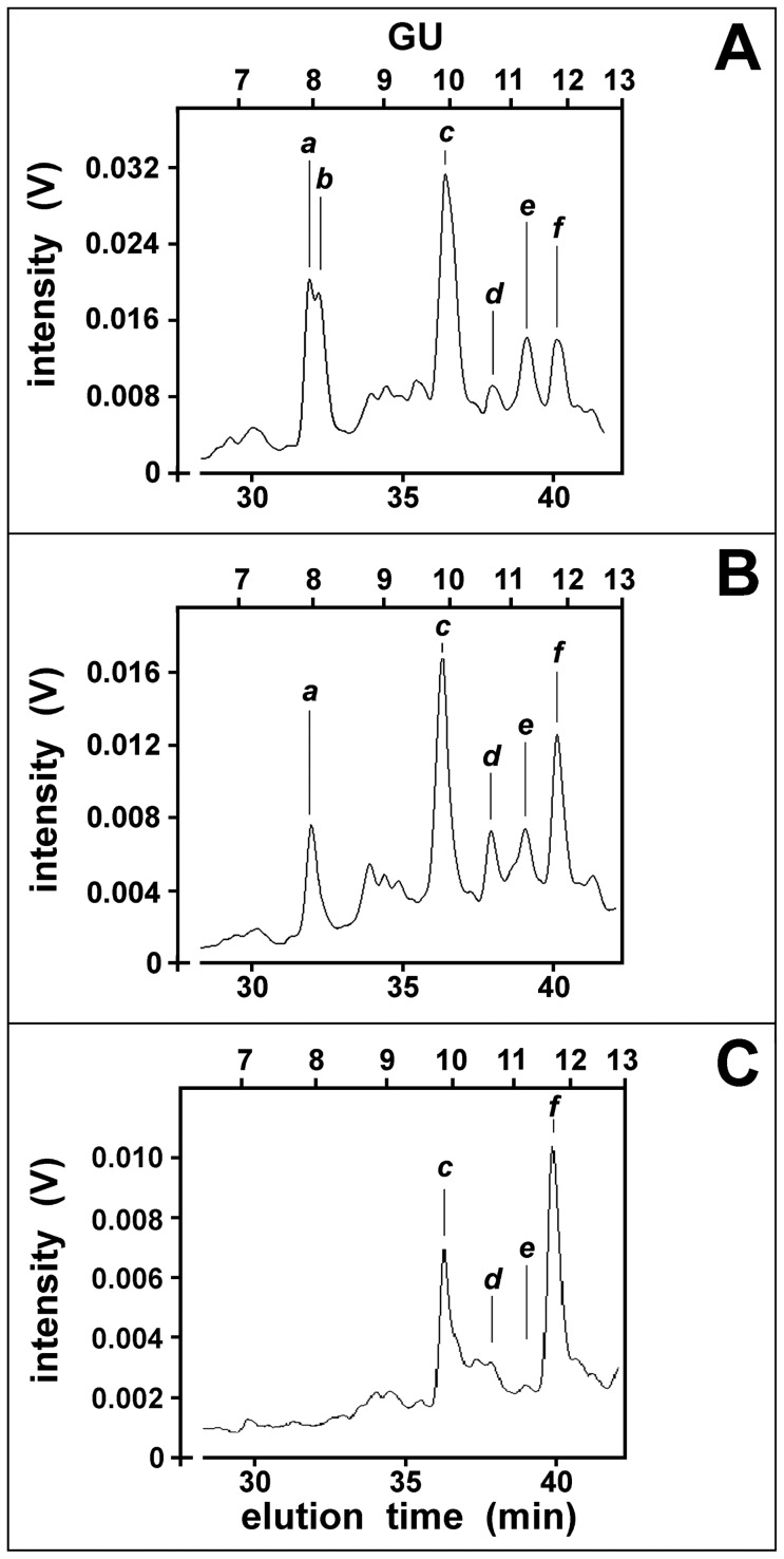
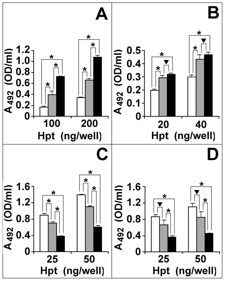
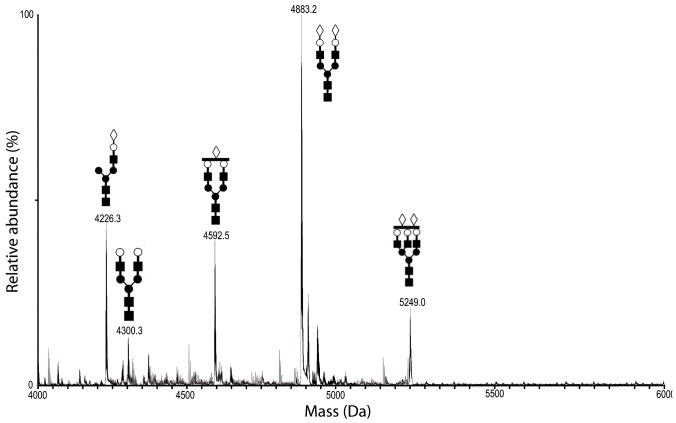
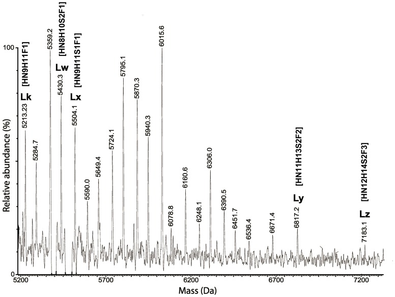
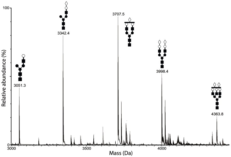
Improved diagnosis of psoriasis, by new biomarkers, is required for evaluating the progression rate of the disease and the response to treatment. Haptoglobin (Hpt), a glycoprotein secreted by hepatocytes and other types of cells including keratinocytes, was found with glycan changes in psoriasis and other diseases. We previously reported that Hpt isolated from plasma of psoriatic patients is more fucosylated than Hpt of healthy subjects. The aim of this study was to compare the glycosylation pattern of Hpt isolated from skin scales or plasma of patients with psoriasis with that of Hpt from cornified epidermal layer or plasma of healthy subjects. High performance liquid chromatography analysis of the glycans isolated from the protein backbone revealed that glycan patterns from skin and plasma of patients were similar, and mostly displayed quantitative rather than qualitative differences from normal pattern. Biotin-labeled lectins were used to evaluate quantitative differences in the glycoforms of Hpt from plasma and psoriatic skin scales. Hpt from skin and plasma of patients showed more fucosylated and branched glycans than Hpt from plasma of healthy subjects. Tryptic glycopeptides of Hpt were also analyzed by mass spectrometry, and a decreased amount of sialylated glycan chains was found in glycopeptides of skin Hpt, as compared with Hpt from plasma. High levels of glycans with fucosylated and tetra-antennary chains were detected on the peptide NLFLNHSENATAK from Hpt of psoriatic patients. Our data demonstrate that specific changes in glycan structures of Hpt, such as enhanced glycan branching and fucose content, are associated with psoriasis, and that differences between circulating and skin Hpt do exist. A lower extent of glycan fucosylation and branching was found in Hpt from plasma of patients in disease remission. Altered glycoforms might reflect changes of Hpt function in the skin, and could be used as markers of the disease.
Conflict of interest statement
Figures
Similar articles
-
Quantitative determination of haptoglobin glycoform variants in psoriasis.Biol Chem. 2010 Dec;391(12):1429-39. doi: 10.1515/BC.2010.146. Biol Chem. 2010. PMID: 21087091
-
Ultrasensitive characterization of site-specific glycosylation of affinity-purified haptoglobin from lung cancer patient plasma using 10 μm i.d. porous layer open tubular liquid chromatography-linear ion trap collision-induced dissociation/electron transfer dissociation mass spectrometry.Anal Chem. 2011 Mar 15;83(6):2029-37. doi: 10.1021/ac102825g. Epub 2011 Feb 21. Anal Chem. 2011. PMID: 21338062 Free PMC article.
-
Glycosylation status of haptoglobin in sera of patients with prostate cancer vs. benign prostate disease or normal subjects.Int J Cancer. 2008 Jan 1;122(1):39-49. doi: 10.1002/ijc.22958. Int J Cancer. 2008. PMID: 17803183
-
Glycobiology of psoriasis: A review.J Autoimmun. 2025 Feb;151:103361. doi: 10.1016/j.jaut.2025.103361. Epub 2025 Jan 13. J Autoimmun. 2025. PMID: 39808852 Review.
-
Overview of Characterizing Cancer Glycans with Lectin-Based Analytical Methods.Methods Mol Biol. 2019;1928:389-408. doi: 10.1007/978-1-4939-9027-6_20. Methods Mol Biol. 2019. PMID: 30725466 Review.
Cited by
-
Haptoglobin as a Biomarker.Biochem Mosc Suppl B Biomed Chem. 2021;15(3):184-198. doi: 10.1134/S1990750821030069. Epub 2021 Aug 16. Biochem Mosc Suppl B Biomed Chem. 2021. PMID: 34422226 Free PMC article.
-
Rapid Evaluation of the Extent of Haptoglobin Glycosylation Using Orthogonal Intact-Mass MS Approaches and Multivariate Analysis.Anal Chem. 2022 Mar 29;94(12):5140-5148. doi: 10.1021/acs.analchem.1c05585. Epub 2022 Mar 14. Anal Chem. 2022. PMID: 35285615 Free PMC article.
-
Comparison of the methods for profiling N-glycans-hepatocellular carcinoma serum glycomics study.RSC Adv. 2018 Jul 20;8(46):26116-26123. doi: 10.1039/c8ra02542h. eCollection 2018 Jul 19. RSC Adv. 2018. PMID: 35541959 Free PMC article.
-
First bioelectronic immunoplatform for quantitative secretomic analysis of total and metastasis-driven glycosylated haptoglobin.Anal Bioanal Chem. 2023 May;415(11):2045-2057. doi: 10.1007/s00216-022-04397-6. Epub 2022 Nov 8. Anal Bioanal Chem. 2023. PMID: 36344668 Free PMC article.
-
Probing N-glycoprotein microheterogeneity by lectin affinity purification-mass spectrometry analysis.Chem Sci. 2019 Apr 16;10(19):5146-5155. doi: 10.1039/c9sc00360f. eCollection 2019 May 21. Chem Sci. 2019. PMID: 31183067 Free PMC article.
References
-
- Langlois MR, Delange JR (1996) Biological and clinical significance of haptoglobin polymorphism in humans. Clin Chem 4: 1589–1600. - PubMed
-
- Wada T, Oara H, Watanabe K, Kinoshita H, Yachi A (1970) Autoradiographic study on the site of uptake of the haptoglobin-hemoglobin complex. J Reticuloendothel Soc 8: 185–193. - PubMed
-
- Bowman BH, Kurosky A (1982) Haptoglobin: the evolutionary product of duplication, unequal crossing over, and point mutation. Adv Hum Genet 12: 189–261, 453–454. - PubMed
-
- Kalmovarin N, Friedrichs WE, O'Brien HV, Linehan LA, Bowman BH, et al. (1991) Extrahepatic expression of plasma protein genes during inflammation. Inflammation 15: 369–379. - PubMed
-
- de Kleijn DP, Smeets MB, Kemmeren PP, Lim SK, Van Middelaar BJ, et al. (2002) Acute-phase protein Haptoglobin is a cell migration factor involved in arterial restructuring. FASEB J 16: 1123–1125. - PubMed
Publication types
MeSH terms
Substances
LinkOut - more resources
Full Text Sources
Other Literature Sources
Medical
