. 2012;7(12):e52065.
doi: 10.1371/journal.pone.0052065. Epub 2012 Dec 18.

Translation directed by hepatitis A virus IRES in the absence of active eIF4F complex and eIF2

Affiliations

Translation directed by hepatitis A virus IRES in the absence of active eIF4F complex and eIF2

Natalia Redondo et al. PLoS One. 2012.

Abstract

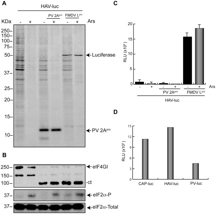
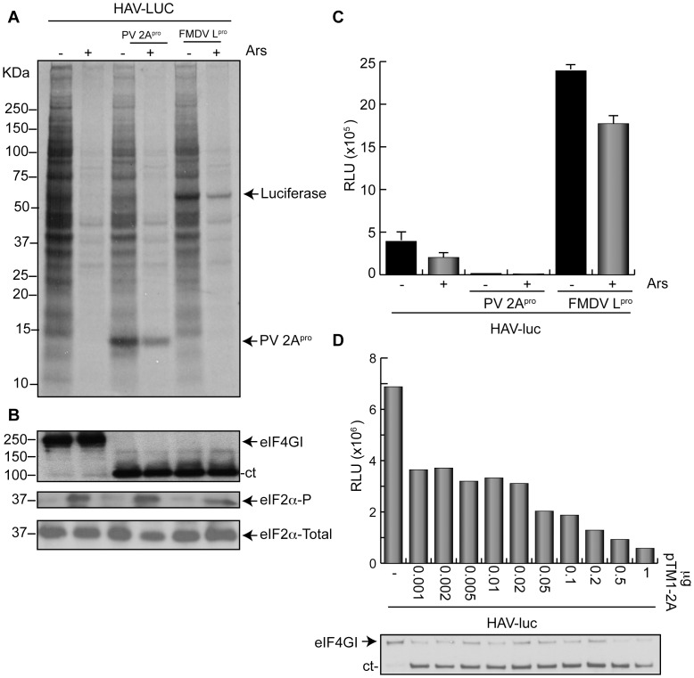
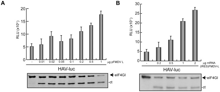
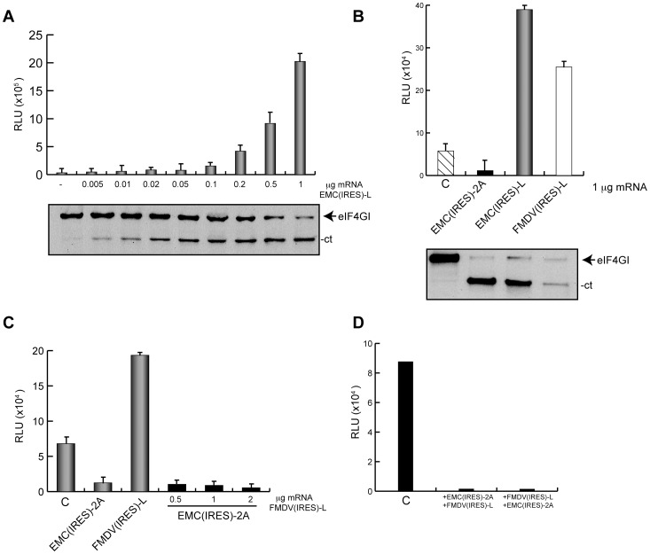
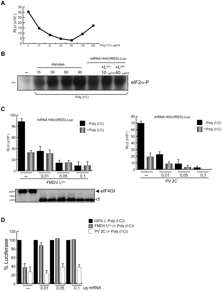
Translation directed by several picornavirus IRES elements can usually take place after cleavage of eIF4G by picornavirus proteases 2A(pro) or L(pro). The hepatitis A virus (HAV) IRES is thought to be an exception to this rule because it requires intact eIF4F complex for translation. In line with previous results we report that poliovirus (PV) 2A(pro) strongly blocks protein synthesis directed by HAV IRES. However, in contrast to previous findings we now demonstrate that eIF4G cleavage by foot-and-mouth disease virus (FMDV) L(pro) strongly stimulates HAV IRES-driven translation. Thus, this is the first observation that 2A(pro) and L(pro) exhibit opposite effects to what was previously thought to be the case in HAV IRES. This effect has been observed both in hamster BHK and human hepatoma Huh7 cells. In addition, this stimulation of translation is also observed in cell free systems after addition of purified L(pro). Notably, in presence of this FMDV protease, translation directed by HAV IRES takes place when eIF2α has been inactivated by phosphorylation. Our present findings clearly demonstrate that protein synthesis directed by HAV IRES can occur when eIF4G has been cleaved and after inactivation of eIF2. Therefore, translation directed by HAV IRES without intact eIF4G and active eIF2 is similar to that observed with other picornavirus IRESs.


Conflict of interest statement

Competing Interests: The authors have declared that no competing interests exist.

Figures

Figure 1
Figure 1. HAV IRES translation in BHK cells after cleavage of eIF4G.
A) BHK-T7 cells were transfected or co-transfected for 2 h with 1 µg plasmid encoding HAV(IRES)-luc alone or in presence of 1 µg pTM1-2A or pFMDV-L, respectively. After 2 hpt, cells were treated with 200 µM Ars for 15 min and then metabolically labeled with 0.2 µCi per well [35S]Met/Cys in presence (+) or absence (−) of Ars for 45 min. Finally, cells were processed by SDS-PAGE, fluorography and autoradiography. B) The same samples were used to analyze eIF4GI, eIF2α phosphorylation and total eIF2α by western blot using specific antibodies as detailed in Materials and Methods. C) BHK-T7 cells were transfected under the conditions described above. Cells were then collected and processed to assay for luc activity as described in Materials and Methods. The bars represent the luc activity in presence (+) or absence (−) of Ars. The RLUs values obtained were as follows: pHAV-luc in absence (−) or presence (+) of Ars were 3.9×10⁵ and 1.8×10⁵, respectively. pHAV-luc co-transfected with pTM1-2A (−) or (+) Ars were 0.2×10⁵ and 0.1×10⁵, respectively, and finally pHAV-luc co-transfected with pFMDV-L (−) or (+) Ars were 25×105 and 17×105, respectively. Error bars indicate standard deviation (SD). D) BHK-T7 cells were transfected with cap-luc, HAV(IRES)-luc or PV(IRES)-luc mRNAs. At 2 hpt cells were collected and luc activity was measured. The RLUs values obtained were as follows: cap-luc: 1.13×10⁶; HAV(IRES)-luc:1.46×10⁶ and PV(IRES)-luc: 0.44×10⁶.
Figure 2
Figure 2. HAV IRES translation in presence of cleaved eIF4G in Huh7-T7 cells.
A) Huh7-T7 cells were transfected or co-transfected for 3 h with 1 µg plasmid encoding HAV(IRES)-luc alone or in presence of 1 µg pTM1-2A or pFMDV-L, respectively. After 2 hpt, cells were treated with 200 µM Ars for 15 min and then metabolically labeled with 0.2 µCi per well[35S]Met/Cys in presence (+) or absence (−) of Ars for 45 min. Finally, cells were processed by SDS-PAGE, fluorography and autoradiography. B) The same samples were used to analyze eIF4GI, eIF2α phosphorylation and total eIF2α by western blot. C) Huh7-T7 cells were transfected under the conditions described above. Cells were then recovered and processed to assay for luc activity as described in Materials and Methods. The bars represent the luc activity in presence (+) or absence (−) of Ars. The RLUs values obtained were as follows: pHAV-luc in absence (−) or presence (+) of Ars were 4.3×10⁵ and 1.8×10⁵, respectively. pHAV-luc co-transfected with pTM1-2A (−) or (+) Ars were 0.3×10⁵ and 0.2×10⁵, respectively, and finally pHAV-luc co-transfected with pFMDV-L (−) or (+) Ars were 23.4×10⁵ and 17.3×10⁵, respectively. Error bars indicate SD. D) Huh7-T7 cells were transfected with 1 µg plasmid pHAV-luc alone or with increasing concentrations of plasmid pTM1-2A for 3 h. After 3 hpt, cells were recovered and processed to measure luc activity. Values obtained are represented in the graph (upper panel). The same samples were used to analyze eIF4GI cleavage (lower panel).
Figure 3
Figure 3. Stimulation of HAV(IRES)-luc mRNA translation is dependent on FMDV Lpro concentration.
A) Huh7-T7 were transfected for 3 h with 1 µg plasmid encoding HAV(IRES)-luc alone and co-transfected with different concentrations of plasmid encoding FMDV Lpro. After 3 hpt, cells were harvested, washed in PBS and resuspended in luc buffer. The graph represents luc synthesis in presence of increasing concentrations of pFMDV-L (upper panel). eIF4GI cleavage was analyzed by western blot (lower panel). B) pFMDV-L was linearized and transcribed in vitro. Huh7-T7 cells were transfected or co-transfected with 1 µg plasmid pHAV-luc alone or with different amounts of FMDV(IRES)-L mRNA. After 3 h in presence of transfection mixture and 3 h in fresh medium, cells were recovered and luc activity was measured and represented in the graph (upper panel). Error bars represent SD. The same samples were employed to analyze eIF4GI cleavage (lower panel).
Figure 4
Figure 4. Co-transfection of HAV(IRES)-luc mRNA and FMDV(IRES)-luc mRNA induces a strong stimulation of luc synthesis.
A) pTM1-L was linearized and transcribed in vitro. EMC(IRES)-L mRNA was obtained. Then, 1 µg pHAV-luc was transfected alone or co-transfected with increasing concentrations of EMC(IRES)-L mRNA for 3 h. At 3 hpt cells were processed as described in Materials and Methods to measure luc activity. Values are represented in the graph (upper panel). Error bars represent SD. The same samples were analyzed by western blot with specific antibodies against eIF4GI (lower panel). B) pHAV-luc was linearized and transcribed in vitro to obtain HAV(IRES)-luc mRNA. Then, 1 µg HAV(IRES)-luc mRNA was transfected alone (C) or co-transfected with 1 µg EMC(IRES)-2A mRNA, 1 µg EMC(IRES)-L or 1 µg FMDV(IRES)-L. At 3 hpt cells were processed to measure luc activity. The values of luc activity are indicated on the graph (upper panel). Error bars represent SD. eIF4GI cleavage was analyzed by western blot (lower panel). C) 1 µg HAV(IRES)-luc mRNA was transfected alone (C) or co-transfected with 1 µg EMC(IRES)-2A mRNA or 1 µg FMDV(IRES)-L mRNA for 3 h. Moreover, an mRNA mixture containing 1 µg HAV(IRES)-luc mRNA, 1 µg EMC(IRES)-2A mRNA and increasing concentrations of FMDV(IRES)-L mRNA were transfected for the same time. At 3 hpt cells were collected and luc activity was measured and plotted. Error bars indicate SD. D) Huh7-T7 cells were transfected with 1 µg HAV(IRES)-luc mRNA alone (C) or co-transfected sequentially with both mRNAs, i.e. first 1 µg EMC(IRES)-2A mRNA or 1 µg FMDV(IRES)-L mRNA was added and incubated for 2 h and then cells were transfected with 1 µg FMDV(IRES)-L mRNA or 1 µg EMC(IRES)-2A mRNA, respectively, together with 1 µg HAV(IRES)-luc. After 2 h of incubation cells were collected and luc activity was measured. The values obtained are represented in the graph.
Figure 5
Figure 5. HAV(IRES)-luc mRNA translation is inhibited by hippuristanol.
A) Effects of hippuristanol in Huh7-T7 cells. Cells were treated for 30 min with increasing concentrations of hippuristanol and then metabolically labelled with 0.2 µCi per well [35S]Met/Cys for 1 h in presence of the inhibitor. Whole-cell extracts were analyzed by SDS-PAGE, fluorography and autoradiography. Dried gels were exposed to X-ray film. B) Huh7-T7 cells were transfected for 3 h with 1 µg plasmid bearing HAV(IRES)-luc in absence (upper panel) or presence (lower panel) of 1 µg pFMDV-L. Then, at 3 hpt, luc activity was measured in presence of increasing concentrations of hippuristanol. C) As control, Huh7-T7 cells were transfected during 3 h with 1 µg pCrPV IGR-luc. After, at 3 hpt, cells were treated with increasing concentrations of hippuristanol for 90 min. Finally, luc activity was measured and the values represented in the graph. Error bars indicate SD.
Figure 6
Figure 6. HAV(IRES)-luc mRNA translation in cell free systems.
A) RRL were incubated with increasing concentrations of poly(I:C) for 30 min at 30°C. After, cap-luc mRNA was added and incubated for 1 h at the same temperature. Then luc activity was measured. The values obtained are represented in the graph. B) RRL were incubated at 30°C for different time periods with 50 ng poly(I:C). In addition, to analyse the effects of mRNA or Lpro on eIF2α phosphorylation, RRL were incubated with the same concentration of poly(I:C) and 100 ng HAV(IRES)-luc mRNA alone or in presence of different amounts of purified Lpro for 30 min at the same temperature. Then, eIF2α phosphorylation was analyzed by western blot. C) Plasmids encoding HAV(IRES)-luc, EMC(IRES)-L and EMC(IRES)-2C were linearized and transcribed in vitro. The translation reaction was then carried out in RRL at 30°C. First, different concentrations of EMC(IRES)-L mRNA was added for 1 h to ensure eIF4G cleavage. Then, the mixture was incubated with 50 ng poly(I:C) during 30 min and finally 100 ng HAV(IRES)-luc mRNA was added and incubated for 1 h at 30°C. As control, EMC(IRES)-2C mRNA was used. In this case, samples were incubated first with different concentrations of EMC(IRES)-2C mRNA. Then, the mixture was incubated with 50 ng poly(I:C) during 30 min and finally, as above, 100 ng HAV(IRES)-luc mRNA was added and incubated for 1 h at 30°C. The graph represents the RLUs from HAV(IRES)-luc mRNA translation in presence of increasing concentrations of EMC(IRES)-L mRNA (left panel) or EMC(IRES)-2C mRNA (right panel). D) Bars represent the percentage of luc synthesis when eIF2α is phosphorylated in the presence of EMC(IRES)-L mRNA or EMC(IRES)-2C mRNA with respect to values without inhibitor, which are taken as 100%.
Figure 7
Figure 7. In vitro translation of HAV(IRES)-luc mRNA in presence of purified Lpro.
HAV IRES was tested in RRL in presence of purified protease FMDV Lpro. First, two different concentrations of protease were added, 10 µg/ml or 40 µg/ml, for 20 min at 30°C. Lysates were then incubated with 50 ng poly(I:C) at the same temperature and,finally, HAV(IRES)-luc mRNA was added and incubated for 1 h at 30°C. Then, aliquots of these samples were processed to measure luc activity (A) and to analyze eIF4GI cleavage (B).

Similar articles

Cited by

References

    1. Belsham GJ (2009) Divergent picornavirus IRES elements. Virus research 139: 183–192. - PubMed
    1. Niepmann M (2009) Internal translation initiation of picornaviruses and hepatitis C virus. Biochimica et biophysica acta 1789: 529–541. - PubMed
    1. Pestova TV, Kolupaeva VG, Lomakin IB, Pilipenko EV, Shatsky IN, et al. (2001) Molecular mechanisms of translation initiation in eukaryotes. Proceedings of the National Academy of Sciences of the United States of America 98: 7029–7036. - PMC - PubMed
    1. Borman AM, Kean KM (1997) Intact eukaryotic initiation factor 4G is required for hepatitis A virus internal initiation of translation. Virology 237: 129–136. - PubMed
    1. Borman AM, Michel YM, Kean KM (2001) Detailed analysis of the requirements of hepatitis A virus internal ribosome entry segment for the eukaryotic initiation factor complex eIF4F. Journal of virology 75: 7864–7871. - PMC - PubMed

Publication types