

Optic nerve magnetisation transfer ratio after acute optic neuritis predicts axonal and visual outcomes
- PMID: 23272235
- PMCID: PMC3525585
- DOI: 10.1371/journal.pone.0052291
Optic nerve magnetisation transfer ratio after acute optic neuritis predicts axonal and visual outcomes
Erratum in
- PLoS One. 2012;7(12). doi: 10.1371/annotation/1c474016-06ef-4827-8fe8-82c158d7616b
Abstract
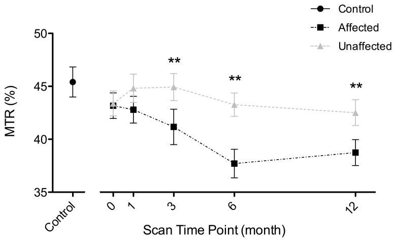
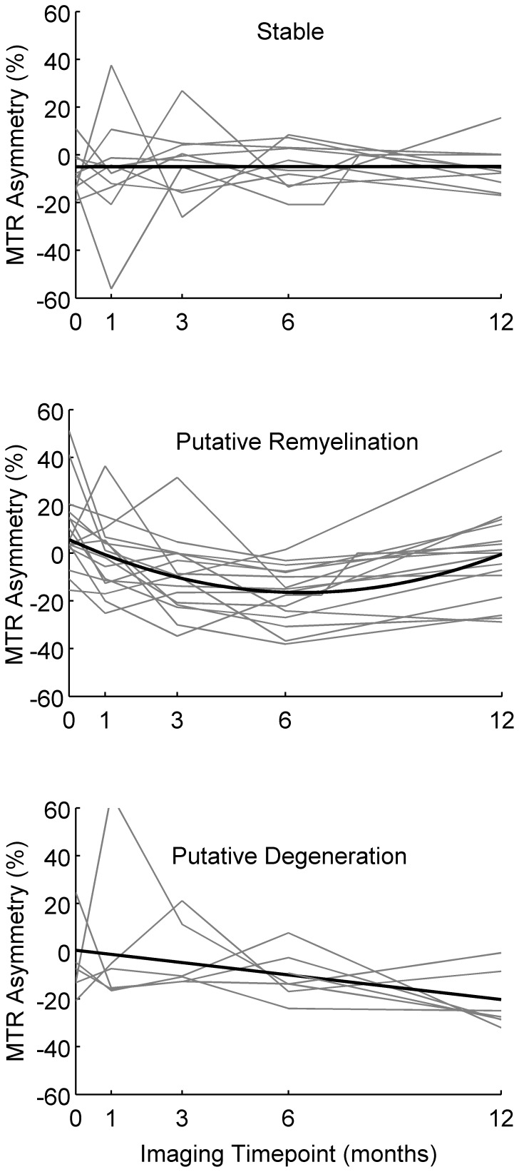
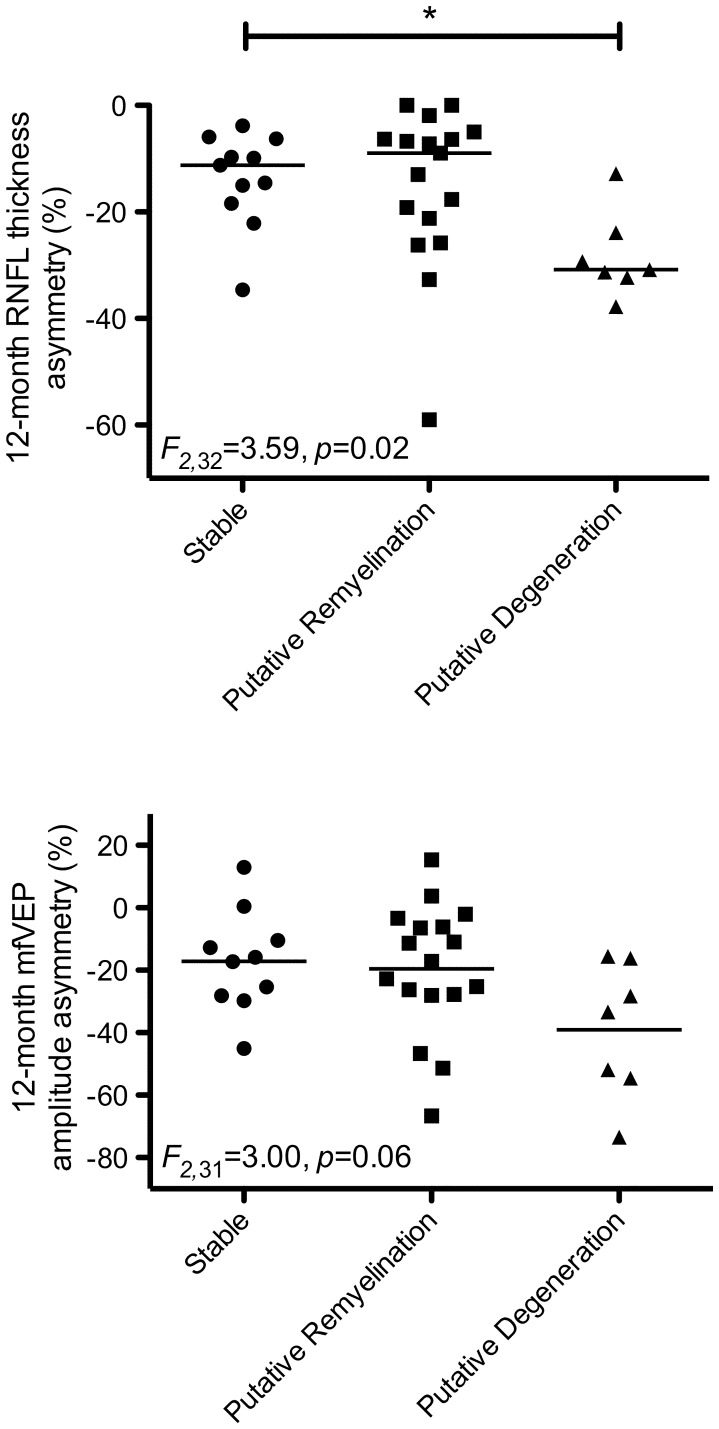
Magnetisation transfer ratio (MTR) can reveal the degree of proton exchange between free water and macromolecules and was suggested to be pathological informative. We aimed to investigate changes in optic nerve MTR over 12 months following acute optic neuritis (ON) and to determine whether MTR measurements can predict clinical and paraclinical outcomes at 6 and 12 months. Thirty-seven patients with acute ON were studied within 2 weeks of presentation and at 1, 3, 6 and 12 months. Assessments included optic nerve MTR, retinal nerve fibre layer (RNFL) thickness, multifocal visual evoked potential (mfVEP) amplitude and latency and high (100%) and low (2.5%) contrast letter acuity. Eleven healthy controls were scanned twice four weeks apart for comparison with patients. Patient unaffected optic nerve MTR did not significantly differ from controls at any time-point. Compared to the unaffected nerve, affected optic nerve MTR was significantly reduced at 3 months (mean percentage interocular difference = -9.24%, p = 0.01), 6 months (mean = -12.48%, p<0.0001) and 12 months (mean = -7.61%, p = 0.003). Greater reduction in MTR at 3 months in patients was associated with subsequent loss of high contrast letter acuity at 6 (ρ = 0.60, p = 0.0003) and 12 (ρ = 0.44, p = 0.009) months, low contrast letter acuity at 6 (ρ = 0.35, p = 0.047) months, and RNFL thinning at 12 (ρ = 0.35, p = 0.044) months. Stratification of individual patient MTR time courses based on flux over 12 months (stable, putative remyelination and putative degeneration) predicted RNFL thinning at 12 months (F(2,32) = 3.59, p = 0.02). In conclusion, these findings indicate that MTR flux after acute ON is predictive of axonal degeneration and visual disability outcomes.
Conflict of interest statement
Figures
Similar articles
-
Optic nerve diffusion tensor imaging after acute optic neuritis predicts axonal and visual outcomes.PLoS One. 2013 Dec 26;8(12):e83825. doi: 10.1371/journal.pone.0083825. eCollection 2013. PLoS One. 2013. PMID: 24386285 Free PMC article.
-
Serial magnetization transfer imaging in acute optic neuritis.Brain. 2004 Mar;127(Pt 3):692-700. doi: 10.1093/brain/awh076. Epub 2004 Jan 21. Brain. 2004. PMID: 14736754
-
Magnetisation transfer ratio in optic neuritis is associated with axonal loss, but not with demyelination.Neuroimage. 2011 May 1;56(1):21-6. doi: 10.1016/j.neuroimage.2011.02.041. Epub 2011 Feb 19. Neuroimage. 2011. PMID: 21338694
-
Assessing structure and function of the afferent visual pathway in multiple sclerosis and associated optic neuritis.J Neurol. 2009 Mar;256(3):305-19. doi: 10.1007/s00415-009-0123-z. Epub 2009 Mar 18. J Neurol. 2009. PMID: 19296047 Review.
-
Visual electrophysiology in the clinical evaluation of optic neuritis, chiasmal tumours, achiasmia, and ocular albinism: an overview.Doc Ophthalmol. 2014 Oct;129(2):71-84. doi: 10.1007/s10633-014-9448-8. Epub 2014 Jun 25. Doc Ophthalmol. 2014. PMID: 24962442 Review.
Cited by
-
Baseline magnetic resonance imaging of the optic nerve provides limited predictive information on short-term recovery after acute optic neuritis.PLoS One. 2015 Jan 30;10(1):e0113961. doi: 10.1371/journal.pone.0113961. eCollection 2015. PLoS One. 2015. PMID: 25635863 Free PMC article.
-
Value of Optic Nerve MRI in Multiple Sclerosis Clinical Management: A MAGNIMS Position Paper and Future Perspectives.Neurology. 2024 Aug 13;103(3):e209677. doi: 10.1212/WNL.0000000000209677. Epub 2024 Jul 17. Neurology. 2024. PMID: 39018513 Free PMC article. Review.
-
Optic nerve diffusion tensor imaging after acute optic neuritis predicts axonal and visual outcomes.PLoS One. 2013 Dec 26;8(12):e83825. doi: 10.1371/journal.pone.0083825. eCollection 2013. PLoS One. 2013. PMID: 24386285 Free PMC article.
-
Monitoring the Course of MS With Optical Coherence Tomography.Curr Treat Options Neurol. 2017 Apr;19(4):15. doi: 10.1007/s11940-017-0452-7. Curr Treat Options Neurol. 2017. PMID: 28374232 Review.
-
Early predictors of visual and axonal outcomes after acute optic neuritis.Front Neurol. 2022 Sep 8;13:945034. doi: 10.3389/fneur.2022.945034. eCollection 2022. Front Neurol. 2022. PMID: 36158958 Free PMC article.
References
-
- Group ONS (1991) The clinical profile of optic neuritis. Experience of the Optic Neuritis Treatment Trial. Arch Ophthalmol 109: 1673–1678. - PubMed
-
- Regan D, Silver R, Murray T (1977) Visual acuity and contrast sensitivity in multiple sclerosis–hidden visual loss: an auxiliary diagnostic test. Brain: a journal of neurology 100: 563. - PubMed
-
- Halliday A, McDonald W, Mushin J (1972) Delayed visual evoked response in optic neuritis. The Lancet 299: 982–985. - PubMed
-
- Costello F, Coupland S, Hodge W, Lorello GR, Koroluk J, et al. (2006) Quantifying axonal loss after optic neuritis with optical coherence tomography. Annals of neurology 59: 963–969. - PubMed
-
- Costello F, Hodge W, Pan Y, Eggenberger E, Coupland S, et al. (2008) Tracking retinal nerve fiber layer loss after optic neuritis: a prospective study using optical coherence tomography. Multiple Sclerosis 14: 893–905. - PubMed
Publication types
MeSH terms
LinkOut - more resources
Full Text Sources
Miscellaneous
