

Analysis of the secretomes of Paracoccidioides mycelia and yeast cells
- PMID: 23272246
- PMCID: PMC3525554
- DOI: 10.1371/journal.pone.0052470
Analysis of the secretomes of Paracoccidioides mycelia and yeast cells
Abstract
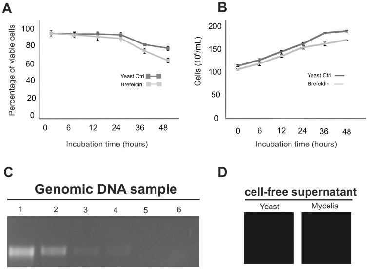
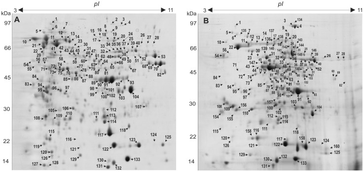
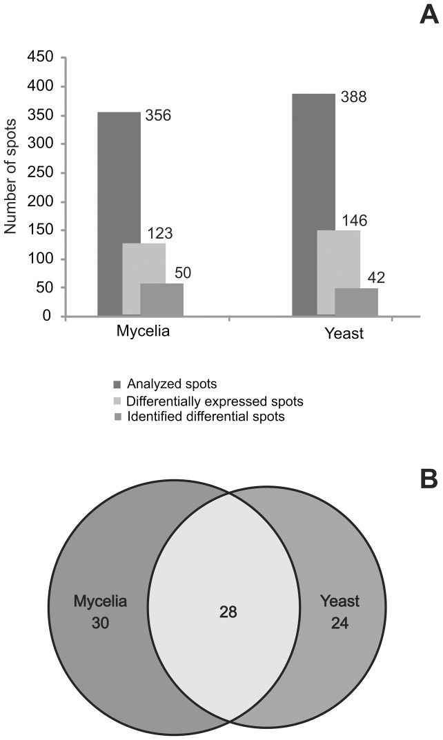
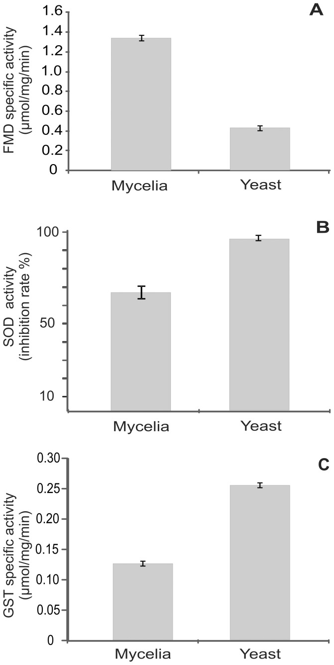
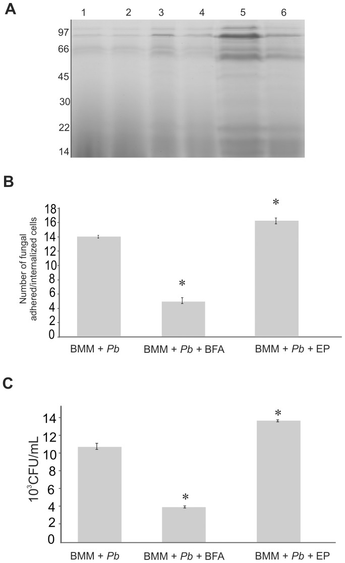
Paracoccidioides, a complex of several phylogenetic species, is the causative agent of paracoccidioidomycosis. The ability of pathogenic fungi to develop a multifaceted response to the wide variety of stressors found in the host environment is important for virulence and pathogenesis. Extracellular proteins represent key mediators of the host-parasite interaction. To analyze the expression profile of the proteins secreted by Paracoccidioides, Pb01 mycelia and yeast cells, we used a proteomics approach combining two-dimensional electrophoresis with matrix-assisted laser desorption ionization quadrupole time-of-flight mass spectrometry (MALDI-Q-TOF MS/MS). From three biological replicates, 356 and 388 spots were detected, in mycelium and yeast cell secretomes, respectively. In this study, 160 non-redundant proteins/isoforms were indentified, including 30 and 24 proteins preferentially secreted in mycelia and yeast cells, respectively. In silico analyses revealed that 65% of the identified proteins/isoforms were secreted primarily via non-conventional pathways. We also investigated the influence of protein export inhibition in the phagocytosis of Paracoccidioides by macrophages. The addition of Brefeldin A to the culture medium significantly decreased the production of secreted proteins by both Paracoccidioides and internalized yeast cells by macrophages. In contrast, the addition of concentrated culture supernatant to the co-cultivation significantly increased the number of internalized yeast cells by macrophages. Importantly, the proteins detected in the fungal secretome were also identified within macrophages. These results indicate that Paracoccidioides extracellular proteins are important for the fungal interaction with the host.
Conflict of interest statement
Figures
Similar articles
-
A quantitative view of the morphological phases of Paracoccidioides brasiliensis using proteomics.J Proteomics. 2011 Dec 21;75(2):572-87. doi: 10.1016/j.jprot.2011.08.020. Epub 2011 Sep 3. J Proteomics. 2011. PMID: 21920475
-
Employing proteomic analysis to compare Paracoccidioides lutzii yeast and mycelium cell wall proteins.Biochim Biophys Acta Proteins Proteom. 2017 Nov;1865(11 Pt A):1304-1314. doi: 10.1016/j.bbapap.2017.08.016. Epub 2017 Aug 24. Biochim Biophys Acta Proteins Proteom. 2017. PMID: 28844734
-
Characterization of extracellular proteins in members of the Paracoccidioides complex.Fungal Biol. 2018 Aug;122(8):738-751. doi: 10.1016/j.funbio.2018.04.001. Epub 2018 Apr 11. Fungal Biol. 2018. PMID: 30007425
-
In vitro, ex vivo and in vivo models: A comparative analysis of Paracoccidioides spp. proteomic studies.Fungal Biol. 2018 Jun;122(6):505-513. doi: 10.1016/j.funbio.2017.10.009. Epub 2017 Oct 28. Fungal Biol. 2018. PMID: 29801795 Review.
-
Functional genome of the human pathogenic fungus Paracoccidioides brasiliensis.FEMS Immunol Med Microbiol. 2005 Sep 1;45(3):369-81. doi: 10.1016/j.femsim.2005.05.013. FEMS Immunol Med Microbiol. 2005. PMID: 16061364 Review.
Cited by
-
Updates in Paracoccidioides Biology and Genetic Advances in Fungus Manipulation.J Fungi (Basel). 2021 Feb 4;7(2):116. doi: 10.3390/jof7020116. J Fungi (Basel). 2021. PMID: 33557381 Free PMC article. Review.
-
Interactome of Glyceraldehyde-3-Phosphate Dehydrogenase Points to the Existence of Metabolons in Paracoccidioides lutzii.Front Microbiol. 2019 Jul 9;10:1537. doi: 10.3389/fmicb.2019.01537. eCollection 2019. Front Microbiol. 2019. PMID: 31338083 Free PMC article.
-
Paracoccidioides Spp.: Virulence Factors and Immune-Evasion Strategies.Mediators Inflamm. 2017;2017:5313691. doi: 10.1155/2017/5313691. Epub 2017 May 2. Mediators Inflamm. 2017. PMID: 28553014 Free PMC article. Review.
-
The Role of the Glutathione System in Stress Adaptation, Morphogenesis and Virulence of Pathogenic Fungi.Int J Mol Sci. 2022 Sep 13;23(18):10645. doi: 10.3390/ijms231810645. Int J Mol Sci. 2022. PMID: 36142553 Free PMC article. Review.
-
The Apoplastic Secretome of Trichoderma virens During Interaction With Maize Roots Shows an Inhibition of Plant Defence and Scavenging Oxidative Stress Secreted Proteins.Front Plant Sci. 2018 Apr 5;9:409. doi: 10.3389/fpls.2018.00409. eCollection 2018. Front Plant Sci. 2018. PMID: 29675028 Free PMC article.
References
-
- Restrepo A, Tobon A (2005) Paracoccdidioides brasiliensis. In: Mandell GL, Bennet JE, Dollin R, editors. Principles and Practice of infectious diseases. Philadelphia. 3062–3068.
-
- Matute DR, McEwen JG, Puccia R, Montes BA, San-Blas G, et al. (2006) Cryptic speciation and recombination in the fungus Paracoccidioides brasiliensis as revealed by gene genealogies. Mol Biol Evol 23: 65–73. - PubMed
-
- Carrero LL, Nino-Vega G, Teixeira MM, Carvalho MJ, Soares CMA, et al. (2008) New Paracoccidioides brasiliensis isolate reveals unexpected genomic variability in this human pathogen. Fungal Genet Biol 45: 605–612. - PubMed
-
- Teixeira MM, Theodoro RC, de Carvalho MJ, Fernandes L, Paes HC, et al. (2009) Phylogenetic analysis reveals a high level of speciation in the Paracoccidioides genus. Mol Phylogenet Evol 52: 273–283. - PubMed
Publication types
MeSH terms
Substances
LinkOut - more resources
Full Text Sources
