Case Reports
. 2012;6(12):e1934.
doi: 10.1371/journal.pntd.0001934. Epub 2012 Dec 13.

Spleen rupture in a case of untreated Plasmodium vivax infection

Affiliations
Case Reports

Spleen rupture in a case of untreated Plasmodium vivax infection

André Machado Siqueira et al. PLoS Negl Trop Dis. 2012.
No abstract available


Conflict of interest statement

The authors have declared that no competing interests exist.

Figures
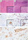
Figure 1
Figure 1. Immunohistochemical staining of P. vivax–infected and normal spleen sections.
HE staining of the normal (A) and P. vivax–infected (B) spleen sections evidencing, respectively, periarteriolar lymphoid cuffs and red pulp cords depleted of lymphoid cells (inset 20× normal) and a prominent white pulp expansion with hyperplastic germinal centers in secondary lymphoid follicles highlighting in the inset a striking red cord infiltration by immunoblasts and reactive plasma cells (20× P. vivax–infected). (C) The normal spleen reveals a scattered CD138 positive plasma cell distribution in the subcapsular and perivascular compartments (2×); higher magnification view shown in the inset (40×). (D) In contrast, a marked increase of CD138 positive plasma cells is observed in the P. vivax–infected spleen (2×), including the detection of mitotic activity among several CD138 positive plasma cells shown in the inset (40×). (E) Double immunostaining with CD138 (brown) and Ki-67 (red) demonstrated the lack of proliferation in plasma cells in the normal spleen (20×). (F) In contrast, there is a significant increase in CD138 and Ki67 positive plasmablasts in the P. vivax–infected spleen (20×).
Figure 2
Figure 2. Immunohistofluorescence images of P. vivax–infected spleen sections.
(A) Spleen section showing P. vivax parasites mostly in the cords of the red pulp as detected by polyclonal antibodies against Vir proteins. (B) Negative control using preimmune sera. Nuclei are shown in blue and the bright field image of the tissue in gray. Scale bar: 20 µm. (C) Double staining showing CD68 macrophages in red and parasites stained in green. Scale bar is 10 µm. (D) Reflection contrast (magenta) was used to detect parasite pigment.

References

    1. Neva FA, Sheagren JN, Shulman NR, Canfield CJ (1970) Malaria: host-defense mechanisms and complications. Ann Intern Med 73: 295–306. - PubMed
    1. Thomas V, Hock SK, Leng YP (1981) Seroepidemiology of malaria: age-specific pattern of Plasmodium falciparum antibody, parasite and spleen rates among children in an endemic area in peninsular Malaysia. Trop Doct 11: 149–154. - PubMed
    1. Brabin L, Brabin BJ, van der Kaay HJ (1988) High and low spleen rates distinguish two populations of women living under the same malaria endemic conditions in Madang, Papua, New Guinea. Trans R Soc Trop Med Hyg 82: 671–676. - PubMed
    1. Hay SI, Smith DL, Snow RW (2008) Measuring malaria endemicity from intense to interrupted transmission. Lancet Infect Dis 8: 369–378. - PMC - PubMed
    1. Chaves LF, Taleo G, Kalkoa M, Kaneko A (2011) Spleen rates in children: an old and new surveillance tool for malaria elimination initiatives in island settings. Trans R Soc Trop Med Hyg 105: 226–231. - PubMed

Publication types