Randomized Controlled Trial
. 2013 Jan;154(1):124-134.
doi: 10.1016/j.pain.2012.09.017.

Amygdala activity contributes to the dissociative effect of cannabis on pain perception

Affiliations
Randomized Controlled Trial

Amygdala activity contributes to the dissociative effect of cannabis on pain perception

Michael C Lee et al. Pain. 2013 Jan.

Abstract

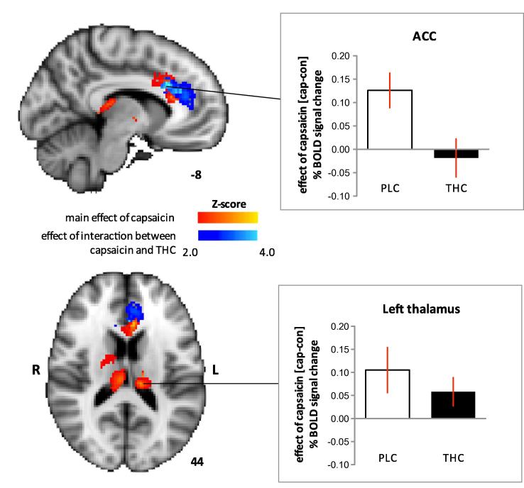
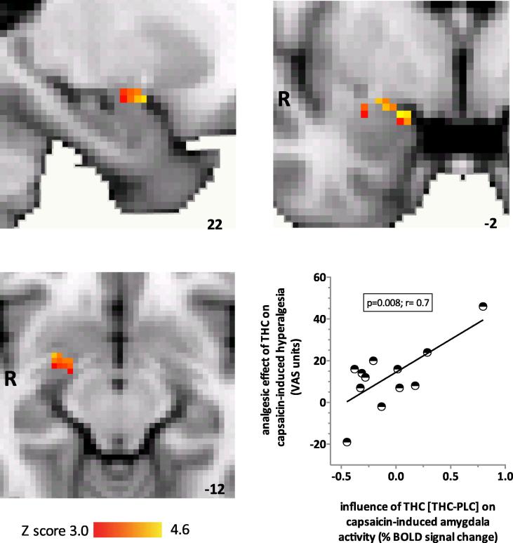
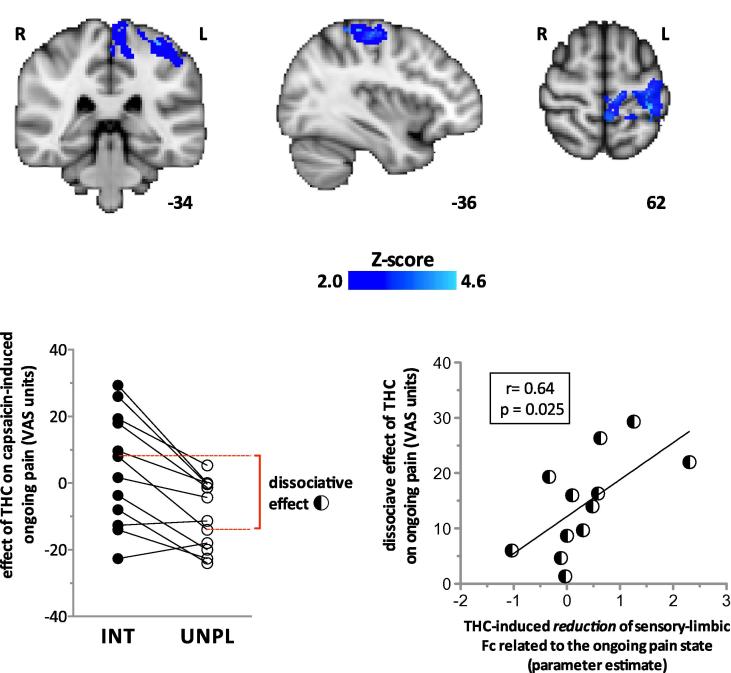
Cannabis is reported to be remarkably effective for the relief of otherwise intractable pain. However, the bases for pain relief afforded by this psychotropic agent are debatable. Nonetheless, the frontal-limbic distribution of cannabinoid receptors in the brain suggests that cannabis may target preferentially the affective qualities of pain. This central mechanism of action may be relevant to cannabinoid analgesia in humans, but has yet to be demonstrated. Here, we employed functional magnetic resonance imaging to investigate the effects of delta-9-tetrahydrocannabinol (THC), a naturally occurring cannabinoid, on brain activity related to cutaneous ongoing pain and hyperalgesia that were temporarily induced by capsaicin in healthy volunteers. On average, THC reduced the reported unpleasantness, but not the intensity of ongoing pain and hyperalgesia: the specific analgesic effect on hyperalgesia was substantiated by diminished activity in the anterior mid cingulate cortex. In individuals, the drug-induced reduction in the unpleasantness of hyperalgesia was positively correlated with right amygdala activity. THC also reduced functional connectivity between the amygdala and primary sensorimotor areas during the ongoing-pain state. Critically, the reduction in sensory-limbic functional connectivity was positively correlated with the difference in drug effects on the unpleasantness and the intensity of ongoing pain. Peripheral mechanisms alone cannot account for the dissociative effects of THC on the pain that was observed. Instead, the data reveal that amygdala activity contributes to interindividual response to cannabinoid analgesia, and suggest that dissociative effects of THC in the brain are relevant to pain relief in humans.


Figures

Fig. 1
Fig. 1
The plasma concentrations of delta-9-tetrahydrocannabinol (THC) and its active metabolite, 11 hydroxy (OH)-THC. Blood was sampled for plasma concentrations about 3.5 hours after THC was given. There were no significant differences (2-tailed paired t test, P > 0.05). Error bars represent SEM. Black and clear bars represent capsaicin and control sessions respectively. n.s., nonsignificant.
Fig. 2
Fig. 2
Capsaicin induced significant ongoing burning pain and caused an increase in pain provoked by punctate stimuli. Compared to placebo, delta-9-tetrahydrocannabinol (THC) significantly reduced the effect of capsaicin on the unpleasantness, but not the intensity of provoked and ongoing pain (Bonferroni adjusted post hoc paired t test, P < 0.05). Error bars represent SEM. Black and clear bars represent THC and placebo, respectively. VAS, visual analogue scale.
Fig. 3
Fig. 3
Delta-9-tetrahydrocannabinol (THC) slowed reaction times (main effect, P < 0.05) and increased resting pulse rate slightly (main effect, P < 0.05). There was no significant effect of capsaicin on reaction times. However, there was a slight increase in resting pulse rate during capsaicin-induced sensitisation compared to control (main effect, P < 0.05). Error bars represent SEM. Black and clear bars represent THC and placebo, respectively.
Fig. 4
Fig. 4
The main effect of capsaicin, indicated in red, was to increase blood-oxygen-level-dependent (BOLD) activation in the anterior cingulate cortex (ACC) (Montreal Neurological Institute [MNI] peak coordinates −6, 20, 30; z score = 3.8) and thalami (left thalamus: MNI peak coordinates −12, −26, 16; z score = 3.2, right thalamus: MNI peak coordinates 10, −22, 14; z score = 3.6). The effect of interaction between delta-9-tetrahydrocannabinol (THC) and capsaicin, indicated in blue, was significant in the ACC only (MNI peak coordinates −8, 22, 28; z score = 4.6). The graphs clarify the effects of THC and placebo (PLC) on capsaicin-induced BOLD responses. Capsaicin-induced BOLD response was calculated as the difference (cap − con) in percentage BOLD signal change between capsaicin (cap) and control (con) sessions. Compared to PLC, THC decreased the BOLD response in the ACC related to hyperalgesia (top graph). In contrast, activation within thalami related to hyperalgesia did not differ significantly. Coloured bars denote range of z scores. Clear and black bars represented PLC and THC, respectively. Error bars represent SEM.
Fig. 5
Fig. 5
Region-of-interest analyses revealed that the main effect of delta-9-tetrahydrocannabinol (THC) was to significantly increase blood-oxygen-level-dependent (BOLD) response within the right amygdala (37 significantly activated voxels; Montreal Neurological Institute [MNI] coordinates of peak voxel 26, 0, −14; z score = 3.23). The coloured bars represent the range of z scores. MNI coordinates are indicated at the bottom right of each slice. The graph shows that in individuals, analgesic effect of THC on the unpleasantness of hyperalgesia was positively and significantly correlated with the effect of THC compared to placebo (PLC) on capsaicin-induced responses in the right amygdala (P < 0.01, r = 0.72). VAS, visual analogue scale.
Fig. 6
Fig. 6
(Top) Delta-9-tetrahydrocannabinol (THC) significantly reduced the right amygdala and primary sensorimotor functional connectivity (Fc) during capsaicin-induced ongoing pain (z > 2.0; cluster-based correction). z-Scores indicating the degree to which THC reduced Fc are scaled in blue. Montreal Neurological Institute (MNI) coordinates are indicated at the bottom right of each slice. (Bottom-left graph) The dissociate effect that THC has on the intensity (INT) and unpleasantness (UNPL) of capsaicin-induced pain in each individual (represented by connecting lines). Negative visual analogue scale (VAS) units on the Y-axis indicate an analgesic effect. The dissociative effect of THC on pain was calculated as the absolute difference of drug effects on the intensity and unpleasantness of ongoing pain, and is indicated in dashed red lines and bracket for an individual. (Bottom-right graph) THC-induced reduction of sensory-limbic Fc was positively correlated with the dissociative effect of the drug on ongoing pain. The interaction term for the drug effect on the sensory-limbic Fc associated with ongoing pain is drug [PLC-THC] × state [CAP-CON]. For each individual, parameter estimates for the interaction effect were calculated for the left-S1 region of interest, which consisted of an 8-mm-diameter sphere centred on the group peak interaction (state × drug) effect within the post-central gyrus (MNI coordinates −34, −36, 62). PLC, placebo; CAP, capsaicin-sensitised state; CON, control state.

References

    1. Abrams D.I., Jay C.A., Shade S.B., Vizoso H., Reda H., Press S., Kelly M.E., Rowbotham M.C., Petersen K.L. Cannabis in painful HIV-associated sensory neuropathy: a randomized placebo-controlled trial. Neurology. 2007;68:515–521. - PubMed
    1. Albe-Fessard D., Berkley K.J., Kruger L., Ralston H.J., III, Willis W.D., Jr. Diencephalic mechanisms of pain sensation. Brain Res. 1985;356:217–296. - PubMed
    1. Amaral D.G., Price J.L. Amygdalo-cortical projections in the monkey (Macaca fascicularis) J Comp Neurol. 1984;230:465–496. - PubMed
    1. Beaulieu P., Ware M. Reassessment of the role of cannabinoids in the management of pain. Curr Opin Anaesthesiol. 2007;20:473–477. - PubMed
    1. Beckmann C.F., Jenkinson M., Smith S.M. General multilevel linear modeling for group analysis in FMRI. Neuroimage. 2003;20:1052–1063. - PubMed

Publication types

MeSH terms