Review
. 2012 Dec 28;13(12):252.
doi: 10.1186/gb-2012-13-12-252.

The ADAR protein family

Review

The ADAR protein family

Yiannis A Savva et al. Genome Biol. .

Abstract

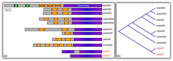
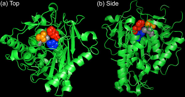
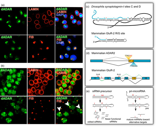
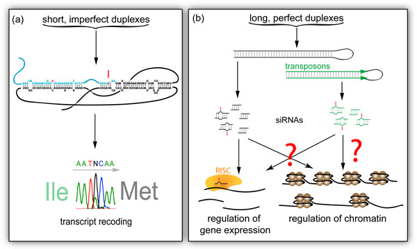
Adenosine to inosine (A-to-I) RNA editing is a post-transcriptional process by which adenosines are selectively converted to inosines in double-stranded RNA (dsRNA) substrates. A highly conserved group of enzymes, the adenosine deaminase acting on RNA (ADAR) family, mediates this reaction. All ADARs share a common domain architecture consisting of a variable number of amino-terminal dsRNA binding domains (dsRBDs) and a carboxy-terminal catalytic deaminase domain. ADAR family members are highly expressed in the metazoan nervous system, where these enzymes predominantly localize to the neuronal nucleus. Once in the nucleus, ADARs participate in the modification of specific adenosines in pre-mRNAs of proteins involved in electrical and chemical neurotransmission, including pre-synaptic release machineries, and voltage- and ligand-gated ion channels. Most RNA editing sites in these nervous system targets result in non-synonymous codon changes in functionally important, usually conserved, residues and RNA editing deficiencies in various model organisms bear out a crucial role for ADARs in nervous system function. Mutation or deletion of ADAR genes results in striking phenotypes, including seizure episodes, extreme uncoordination, and neurodegeneration. Not only does the process of RNA editing alter important nervous system peptides, but ADARs also regulate gene expression through modification of dsRNA substrates that enter the RNA interference (RNAi) pathway and may then act at the chromatin level. Here, we present a review on the current knowledge regarding the ADAR protein family, including evolutionary history, key structural features, localization, function and mechanism.


Figures

Figure 1
Figure 1
The ADAR family protein. (a) Domain architecture of metazoan ADARs. The deaminase domain is depicted in purple, while the dsRBMs are shown in orange and Z-DNA binding domains, unique to human ADAR1, are presented in green. The human genome contains three ADAR genes (hADAR1 to 3). That of the squid Loligo pealeii contains an ADAR2-like gene (sqADAR2) that produces variants (a and b) through alternative splicing. C. elegans contains two genes (ceADAR1 and 2), while the genome of D. melanogaster encodes only one (dADAR), an enzyme homologous to hADAR2. Although the dsRBMs found in the Hydra magnapapillata genome are highly divergent, five such motifs are recognizable in hmADAR, the only identified gene in this species. Human and Drosophila ADAT architectures are included (red), as these enzymes are believed to be ancestral to present-day ADARs. (b) Cladogram based on ADAR catalytic domain sequences. MacVector was used to generate a relatedness tree based on the protein sequences of ADAR catalytic domains from different species. C. elegans ADAR2 is absent due to difficulty aligning the catalytic domain. Note that human and Drosophila ADATs (red) cluster as the outgroup.
Figure 2
Figure 2
Crystal structure of the human ADAR2 deaminase domain shown from the top (a) and side (b). The catalytic core is formed between H394 (red), E396 (blue), C451 (orange) and C516 (gray). The core also includes a water molecule, zinc ion and IP6 molecule (not shown).
Figure 3
Figure 3
Overview of ADAR localization and function. (a) Transgenic HA-tagged ADAR (green) localizes within the nuclear envelope (lamin, red) and more specifically to the nucleolus (fibrillarin, red) in Drosophila salivary gland cells. (b) Endogenous HA-tagged ADAR (green) localizes to the Drosophila neuronal nucleus and colocalizes with the nucleolus, distinguished by the red fibrillarin signal (arrowheads). (c) RNA structures that direct editing. The complex pseudoknot of Drosophila synaptotagmin-I is presented in contrast with the simple hairpin of mammalian GluR-2, both specific editing targets. Exons are represented in blue, introns in black. Adenosines targeted by ADAR are red. (d) Editing affects splicing. Mammalian ADAR2 auto-edits its own transcript, creating a novel splice site (red), which results in the inclusion of 47 nucleotides (yellow) and a frameshift in the coding sequence. In the mammalian GluR-2 transcript, editing at both the Q/R site in exon 11 and an intronic 'hotspot' (red) is required for efficient removal of the downstream intron. Editing of the R/G site (red) reduces efficacy of downstream splicing and favors an alternative final exon configuration (yellow). (e) Editing can interfere with siRNA and microRNA production and targeting. Perfectly duplex siRNA precursors are targets for hyper-editing by ADARs. Editing may result in improper Dicer processing and fewer functional siRNAs, or edited siRNAs. Primary (pri) microRNAs, imperfect duplexes, may be targets for specific editing, resulting in mature miRNAs toward alternative mRNA targets.
Figure 4
Figure 4
The consequences of specific versus non-specific editing. (a) Short, imperfect duplexes, such as Drosophila synaptotagmin-I, are specifically edited leading to transcript and peptide recoding. (b) Long, perfect dsRNA substrates, including those formed by nearby transposons in opposite orientation (green), are hyper-edited, leading to fewer or edited siRNAs. This may alter gene expression through RISC (yellow) targeting, but evidence also links the RNAi pathway to chromatin regulation.

References

    1. Basillo C, Wahba A, Lengyel P, Speyer J, Ochoa S. Synthetic polynucleotides and the amino acid code. V Proc Natl Acad Sci USA. 1962;48:613–616. doi: 10.1073/pnas.48.4.613. - DOI - PMC - PubMed
    1. Bass BL, Weintraub H. A developmentally regulated activity that unwinds RNA duplexes. Cell. 1987;48:607–613. doi: 10.1016/0092-8674(87)90239-X. - DOI - PubMed
    1. Bass BL. RNA editing by adenosine deaminases that act on RNA. Annu Rev Biochem. 2002;71:817–846. doi: 10.1146/annurev.biochem.71.110601.135501. - DOI - PMC - PubMed
    1. Nishikura K. Functions and regulation of RNA editing by ADAR deaminases. Annu Rev Biochem. 2010;79:321–349. doi: 10.1146/annurev-biochem-060208-105251. - DOI - PMC - PubMed
    1. Chen CX, Cho DS, Wang Q, Lai F, Carter KC, Nishikura K. A third member of the RNA-specific adenosine deaminase gene family, ADAR3, contains both single- and double-stranded RNA binding domains. RNA. 2000;6:755–767. doi: 10.1017/S1355838200000170. - DOI - PMC - PubMed

Substances