

Synaptic genes are extensively downregulated across multiple brain regions in normal human aging and Alzheimer's disease
- PMID: 23273601
- PMCID: PMC4022280
- DOI: 10.1016/j.neurobiolaging.2012.11.024
Synaptic genes are extensively downregulated across multiple brain regions in normal human aging and Alzheimer's disease
Abstract
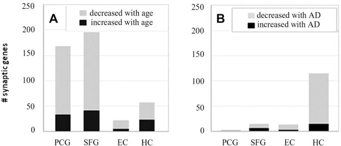
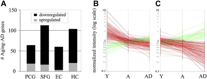
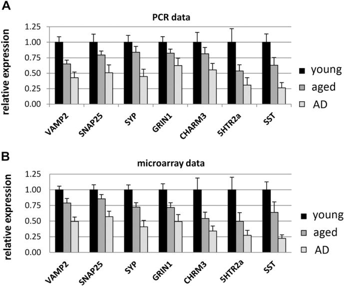
Synapses are essential for transmitting, processing, and storing information, all of which decline in aging and Alzheimer's disease (AD). Because synapse loss only partially accounts for the cognitive declines seen in aging and AD, we hypothesized that existing synapses might undergo molecular changes that reduce their functional capacity. Microarrays were used to evaluate expression profiles of 340 synaptic genes in aging (20-99 years) and AD across 4 brain regions from 81 cases. The analysis revealed an unexpectedly large number of significant expression changes in synapse-related genes in aging, with many undergoing progressive downregulation across aging and AD. Functional classification of the genes showing altered expression revealed that multiple aspects of synaptic function are affected, notably synaptic vesicle trafficking and release, neurotransmitter receptors and receptor trafficking, postsynaptic density scaffolding, cell adhesion regulating synaptic stability, and neuromodulatory systems. The widespread declines in synaptic gene expression in normal aging suggests that function of existing synapses might be impaired, and that a common set of synaptic genes are vulnerable to change in aging and AD.
Published by Elsevier Inc.
Conflict of interest statement
The authors have no actual or potential conflicts of interest.
All case identifiers have been removed and replaced with a unique case number by the providing tissue repositories, in compliance with protection of subjects and institutional review board (IRB) requirements.
Figures
References
-
- Adams I. Comparison of synaptic changes in the precentral and postcentral cerebral cortex of aging humans: a quantitative ultrastructural study. Neurobiol Aging. 1987;8:203–212. - PubMed
-
- Bannerman DM. Fractionating spatial memory with glutamate receptor subunit-knockout mice. Biochem Soc Trans. 2009;37:1323–1327. - PubMed
-
- Braak H, Braak E. Neuropathological stageing of Alzheimer-related changes. Acta Neuropathol. 1991;82:239–259. - PubMed
Publication types
MeSH terms
Grants and funding
- AG34667/AG/NIA NIH HHS/United States
- AG7367/AG/NIA NIH HHS/United States
- R01 AG23173/AG/NIA NIH HHS/United States
- R01 AG36400/AG/NIA NIH HHS/United States
- R01 AG034667/AG/NIA NIH HHS/United States
- P50 AG16573/AG/NIA NIH HHS/United States
- R56 AG007367/AG/NIA NIH HHS/United States
- P50 AG016573/AG/NIA NIH HHS/United States
- AG00538/AG/NIA NIH HHS/United States
- R01 AG036400/AG/NIA NIH HHS/United States
- R01 AG007367/AG/NIA NIH HHS/United States
- P01 AG000538/AG/NIA NIH HHS/United States
- R01 AG023173/AG/NIA NIH HHS/United States
LinkOut - more resources
Full Text Sources
Other Literature Sources
Medical
Molecular Biology Databases
