

Comment
doi: 10.1016/j.molcel.2012.12.008.
A dancer caught midstep: the structure of ATP-bound Hsp70
Affiliations
- PMID: 23273742
- PMCID: PMC3746503
- DOI: 10.1016/j.molcel.2012.12.008
Item in Clipboard
Comment
A dancer caught midstep: the structure of ATP-bound Hsp70
Mol Cell.
.
Abstract
Hsp70 ATP binding induces substrate release, but the transiency of this state has inhibited its characterization. In this issue, Kityk et al. determine the Hsp70(∗)ATP structure utilizing engineered disulfide bonds, providing insights into the workings of this essential molecular machine.
Copyright © 2012 Elsevier Inc. All rights reserved.
Figures
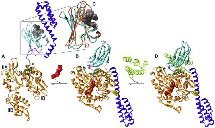
(A) In the ADP/apo state, substrates (gray) are bound within the SBD β sandwich (cyan) and capped by the SBD helical lid (blue; Protein Data Bak [PDB] 2KHO [Bertelsen et al., 2009] with the substrate imported from PDB 1DKX [Zhu et al., 1996]). (B) Upon binding ATP (red), the NBD (orange) closes, leading to widening of the crevice between subdomains 1A and IIA, allowing the NBD-SBD linker (magenta) to bind within it. Together with other changes in the disposition of NBD regions (in green) involved in interactions with SBDβ, this creates a surface on which SBDβ docks, leading to displacement of SBDα, which docks onto subdomain 1B (PDB 4B9Q [Kityk et al., 2012]; cysteines introduced to create a disulfide to lock in this conformation for crystallization are in yellow). (C) Closed SBDβ (orange) wraps around bound substrates but is induced to open (cyan) and release substrate by interactions with NBD*ATP. Substrate rebinding induces SBDβ closure and transmits an ATPase stimulating signal to the NBD through the NBD:SBDβ interface. (D) The ATPase stimulating signal is amplified by the J cochaperone (in light green and imported from PDB 2QWN [Jiang et al., 2007]), which binds so as to hold SBDβ and the linker against the NBD.
Comment on
-
Structure and dynamics of the ATP-bound open conformation of Hsp70 chaperones.Mol Cell. 2012 Dec 28;48(6):863-74. doi: 10.1016/j.molcel.2012.09.023. Epub 2012 Nov 1. Mol Cell. 2012. PMID: 23123194
References
Publication types
Grants and funding
LinkOut - more resources
Full Text Sources
