Review
. 2013:300:1-50.
doi: 10.1016/B978-0-12-405210-9.00001-1.

New insights into mechanisms of stem cell daughter fate determination in regenerative tissues

Affiliations
Review

New insights into mechanisms of stem cell daughter fate determination in regenerative tissues

Aiko Sada et al. Int Rev Cell Mol Biol. 2013.

Abstract

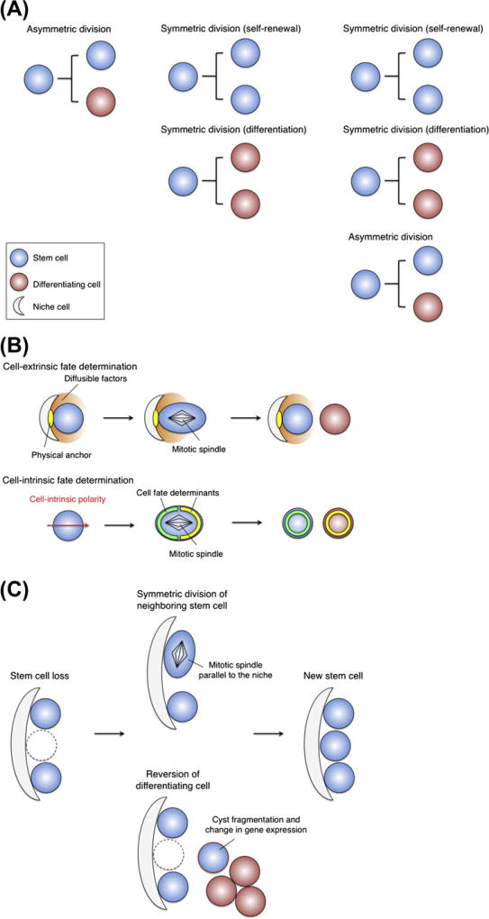
Stem cells can self-renew and differentiate over extended periods of time. Understanding how stem cells acquire their fates is a central question in stem cell biology. Early work in Drosophila germ line and neuroblast showed that fate choice is achieved by strict asymmetric divisions that can generate each time one stem and one differentiated cell. More recent work suggests that during homeostasis, some stem cells can divide symmetrically to generate two differentiated cells or two identical stem cells to compensate for stem cell loss that occurred by direct differentiation or apoptosis. The interplay of all these factors ensures constant tissue regeneration and the maintenance of stem cell pool size. This interplay can be modeled as a population-deterministic dynamics that, at least in some systems, may be described as stochastic behavior. Here, we overview recent progress made on the characterization of stem cell dynamics in regenerative tissues.


Figures

Figure 1
Figure 1
Stem cell behavior proposed in invertebrate model systems. (A) Three possible cell division strategies: invariant asymmetric division (left); invariant symmetric division (middle); combination of asymmetric and symmetric divisions (right). (B) Cell-extrinsic (upper) and -intrinsic (lower) regulation of asymmetric cell division. (C) Two possible stem cell behaviors to replenish a new stem cell: symmetric division (upper) and dedifferentiation (lower).
Figure 2
Figure 2
H2B-GFP tet-off system to count cell division frequency in vivo. (A) Schematic representation of the strategy to detect slow-cycling cells with H2B-GFP. Doxy administration inhibits the binding of tetR-VP16 proteins to the TRE promoter, and thus turns off H2B-GFP transcription. Cells dilute H2B-GFP proteins after division, which enables quantification of the frequency of cell division during chase periods. (B) H2B-GFP pulse-chase in skin (Tumbar et al., 2004; Waghmare et al., 2008). The keratin5 promoter drives H2B-GFP expression in skin epithelial cells. After 3 weeks of doxy chase, bulge contains cells with different cell-division history shown in FACS profile on the right.
Figure 3
Figure 3
Genetic lineage-tracing experiments. (A) Schematic representation of the double transgenic DNA construct used for lineage-tracing experiments. Tamoxifen administration translocates CreER to the nucleus, where Cre-mediated recombination takes place. (B) Example of lineage-tracing experiments using Tamoxifen-inducible Cre driven by spermatogonia-specific Nanos2 enhancer/promoter (Sada et al., 2009). Two days after Tamoxifen injection, spermatogonia located on the basement membrane are labeled. Three months after labeling, a sufficiently long period for a repeated completion of spermatogenesis, seminiferous tubule of testis contained all stages of spermatogenic germ cells.
Figure 4
Figure 4
Interfollicular epidermis. (A) EPU model. A slow-cycling stem cell lies at the center of each unit and generates TA cells. Postmitotic basal cells leave the basal layer and migrate vertically to the suprabasal layer. (B) Distribution of epidermal clone. In EPU model, the size and shape of the labeled clones is constant (left). In actual observation, the clone size increases with time (right). (C) New model proposes three outcomes of the committed progenitor (CP) cell division.
Figure 5
Figure 5
Hair follicle stem cells. (A) The hair follicle structure. (B) The hair follicle cycle: stages of rest (telogen), growth (anagen), regression (catagen), and a less synchronous stage of hair shedding (exogen). (C) Models for symmetric fate decisions for hair follicle stem cells during hair cycle.
Figure 6
Figure 6
Murine spermatogonial stem cells. (A) A testis is composed of long, coiled tubes called seminiferous tubules. Spermatogonia are located on the basement membrane of seminiferous tubules. As germ cells mature, they progressively locate toward the lumen of the tubules. Sertoli cells enclose germ cells within tubules, while vascular and surrounding interstitial cells are located outside of the tubules. Spermatogonia are interconnected by intercellular bridges and classified by the number of cell(s) in the same cluster. (B) Clonal analysis in the mouse testis. The number of labeled clones per testis decreases with time, while the average clone length increases. (C) Interpretation of clone expansion or loss. The clone expansion is caused by a loss of unlabeled stem cell and a subsequent replacement by labeled stem cell (upper), whereas the clone loss occurs by the opposite labeling pattern of stem cells (lower).
Figure 7
Figure 7
Intestinal stem cells. (A) The anatomy of the small intestinal epithelium. (B) Confetti reporter construct. Cre triggers both inversion and recombination in a random manner, which results in the four patterns of gene expression (nuclear GFP, cytoplasmic YFP and RFP, membrane-associated CFP). Cre-mediated inversion occurs at a sequence flanked by loxP sites in opposite orientation. In 50% of cells, inversion should lead to an antisense orientation and switch gene expression. (C) Multicolor lineage tracing shows a progressive monoclonality of the crypt. (D) Monoclonal conversion arises from turnover of an equipotent stem cell population. Paneth cells make a specialized microenvironment for intestinal stem cells.

References

    1. Ahmed S. The culture of neural stem cells. J. Cell. Biochem. 2009;106:1–6. - PubMed
    1. Altman J, Das GD. Autoradiographic and histological evidence of postnatal hippocampal neurogenesis in rats. J. Comp. Neurol. 1965;124:319–335. - PubMed
    1. Ambler CA, Maatta A. Epidermal stem cells: location, potential and contribution to cancer. J. Pathol. 2009;217:206–216. - PubMed
    1. Bardin AJ, et al. Asymmetric localization and function of cell-fate determinants: a fly’s view. Curr. Opin. Neurobiol. 2004;14:6–14. - PubMed
    1. Barker N, et al. Identification of stem cells in small intestine and colon by marker gene Lgr5. Nature. 2007;449:1003–1007. - PubMed

Publication types

LinkOut - more resources