. 2013 Jan 31;3(1):30-5.
doi: 10.1016/j.celrep.2012.12.001. Epub 2012 Dec 27.

Structure of a pestivirus envelope glycoprotein E2 clarifies its role in cell entry

Affiliations

Structure of a pestivirus envelope glycoprotein E2 clarifies its role in cell entry

Kamel El Omari et al. Cell Rep. .

Abstract

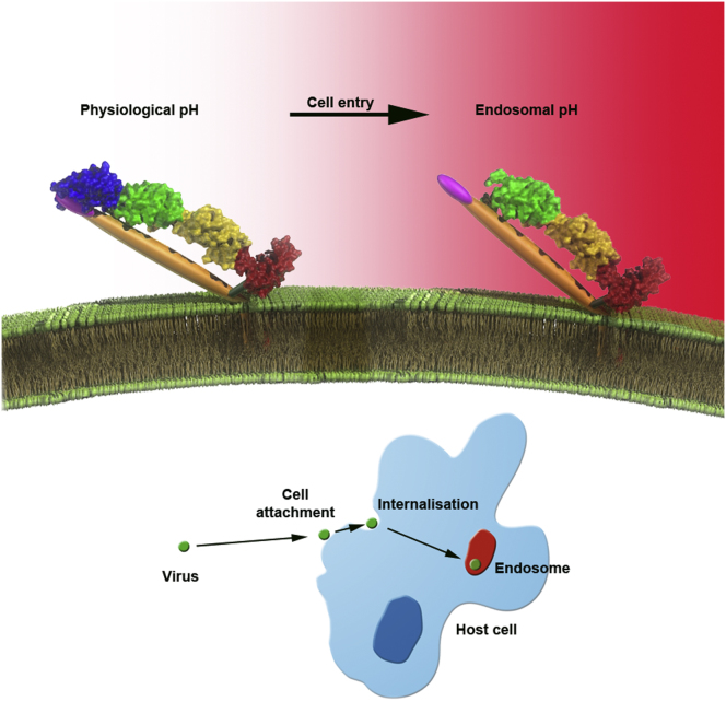
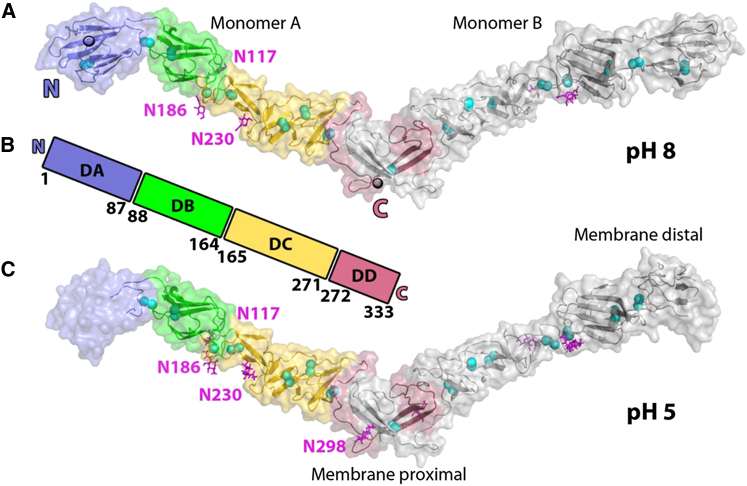
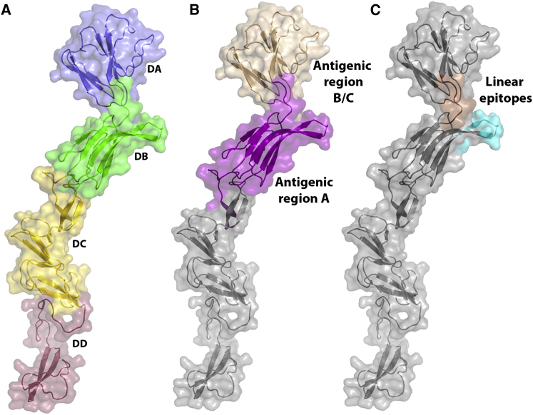
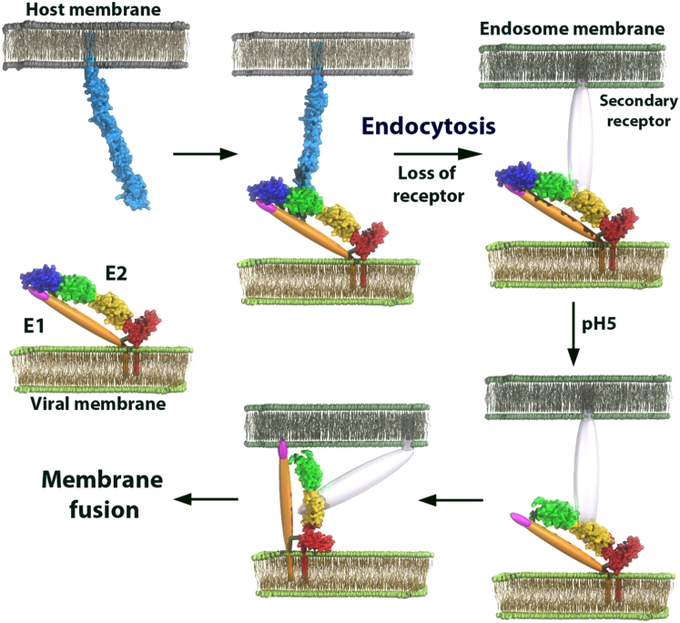
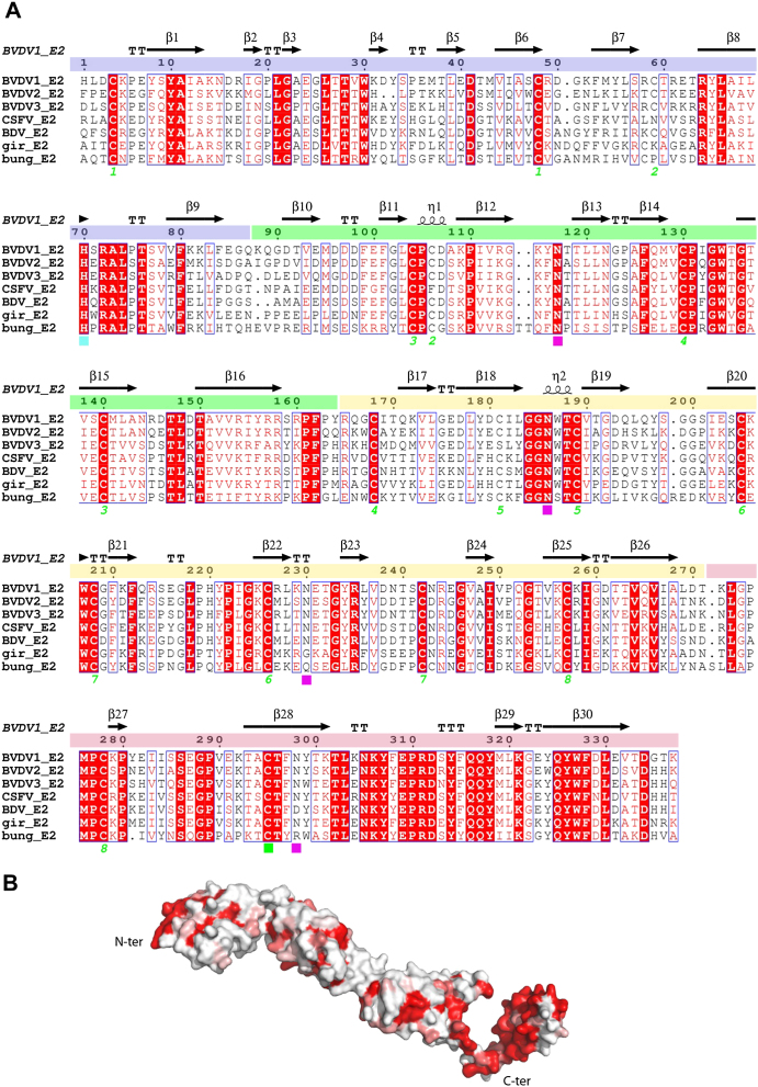
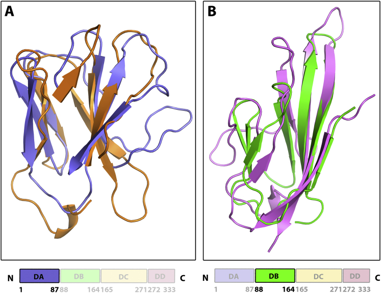
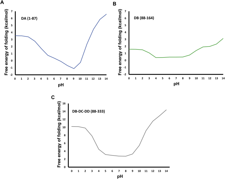
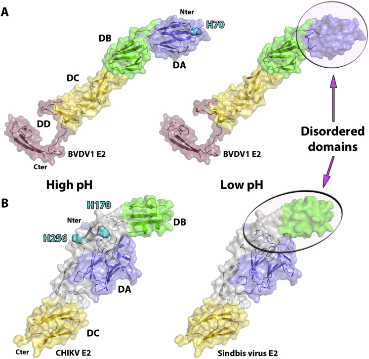
Enveloped viruses have developed various adroit mechanisms to invade their host cells. This process requires one or more viral envelope glycoprotein to achieve cell attachment and membrane fusion. Members of the Flaviviridae such as flaviviruses possess only one envelope glycoprotein, E, whereas pestiviruses and hepacivirus encode two glycoproteins, E1 and E2. Although E2 is involved in cell attachment, it has been unclear which protein is responsible for membrane fusion. We report the crystal structures of the homodimeric glycoprotein E2 from the pestivirus bovine viral diarrhea virus 1 (BVDV1) at both neutral and low pH. Unexpectedly, BVDV1 E2 does not have a class II fusion protein fold, and at low pH the N-terminal domain is disordered, similarly to the intermediate postfusion state of E2 from sindbis virus, an alphavirus. Our results suggest that the pestivirus and possibly the hepacivirus fusion machinery are unlike any previously observed.


Figures

None
Graphical abstract
Figure 1
Figure 1
Overall Fold of BVDV1 E2 (A) Cartoon representation of the crystal structure of BVDV1 E2 obtained at pH8. A dimer of BVDV1 E2 is seen in the crystal. Domains of monomer A starting from the N terminus are colored in blue, green, yellow and red, respectively. The sugars are shown as magenta sticks and disulfide bridges are represented as cyan spheres. (B) Linear representation of BVDV1 E2 showing the four domains DA, DB, DC and DD with their domain boundaries. Coloring as in a. (C) Cartoon representation of the crystal structure of BVDV1 E2 obtained at pH5. Domains DA of monomers A and B are disordered (the transparent surface rendered shows the position of the domains when ordered at higher pH). See also Figures S1, S2, S3, and S4.
Figure 2
Figure 2
Mapping of Epitopes of Classical Swine Fever Virus E2 onto the Structure of BVDV E2 (A) Cartoon representation of a monomer of BVDV1 E2 colored by domain as in Figure 1. (B) Positions of the antigenic regions A and B/C. (C) Location of the linear epitopes LFDGTNP (orange) and TAVSPTTLR (cyan). LFDGTNP is at the border of the two antigenic regions, whereas TAVSPTTLR is located in the antigenic region A. See also Figure S1.
Figure 3
Figure 3
Interactions of BVDV1 E2 and Their Locations (A) Location of histidine (H70) of domain DA, which is conserved among pestiviruses and that may trigger an order-disorder transition of this domain at low pH. (B) The potential host cell receptor binding site in domain DB colored in pink. Domain DB is rotated as shown. (C) The interface of monomers A (shown colored) and B (shown in gray) contains a strand swap and hydrophobic residues such as phenylalanines, tyrosines and tryptophanes drawn as sticks. Domains DD of molecules A and B are linked by a disulfide bridge (cyan sphere) to form a covalent homodimer. See also Figure S1.
Figure 4
Figure 4
Proposed Model of the Fusion Mechanism of Pestiviruses A covalently linked E1-E2 heterodimer is present on the viral membrane. E2 binds to the host receptor (possibly CD46, shown in light blue) through domain DB prior to endocytosis. As CD46 is not internalized, a putative secondary receptor (transparent) presumably replaces CD46. Throughout this process the putative fusion loop of E1 (magenta), is concealed by domain DA of E2. At low pH, domain DA of E2 becomes disordered and exposes the fusion loop that will then be able to insert into the endosome membrane and trigger membrane fusion. It is as yet unknown whether E1 forms trimers as seen for other fusion proteins.
Figure S1
Figure S1
Amino Acid Conservation between Pestiviruses E2 Glycoprotein, Related to Figures 1, 2, and 3 (A) Sequence alignment of BVDV1 E2 with E2 proteins from other pestiviruses: BVDV2, BVDV3, Classical Swine Fever virus (CSFV), Border Disease virus (BDV), Pestivirus of giraffe (gir) and Bungowannah virus (bung). Conserved residues are drawn in red boxes, similar residues in red type. The secondary structure assignment for BVDV1 E2 is shown at the top and domains DA, DB, DC and DD are colored in blue, green, yellow and red, respectively. Cysteines involved in intra-chain disulfide bridges are marked by a green number, the single cysteine involved in the inter-chain disulfide bridge is marked by a green square, the four glycosylation sites of BVDV1 E2 are marked with magenta squares and the conserved histidine is marked with a cyan square. (B) Surface representation of a monomer of BVDV E2 colored according to sequence conservation from white (non-conserved) to red (conserved).
Figure S2
Figure S2
Domains DA and DB Have Ig-fold, Related to Figure 1 (A) Domain DA of BVDV1 E2 (neutral pH form) is colored in blue and is superimposed on a T cell receptor alpha chain (PDB: 2Z31, Z = 5.3, rmsd = 2.8Å) colored in orange. (B) Domain DB is colored in green and is superimposed on the C-terminal domain of semaphorin 4D (PDB: 1OLZ, Z = 6.7, rmsd = 2.3Å) colored in pink.
Figure S3
Figure S3
Stability of BVDV1 E2 Domains over pH, Related to Figure 1 (A) domain DA stability depends on pH, the results would suggest that domain become less stable when moving from physiological pH (7.4) to the endosome pH 5. (B) For comparison, domain DB, a similarly sized Ig-like domain to domain DA, would not be destabilized by a change of pH from 7.4 to 5. (C) Domains DB-DC-DD would not be destabilized by a change of pH from 7.4 to 5.
Figure S4
Figure S4
Structural Comparison of Pestivirus and Alphavirus E2 Glycoproteins at High and Low pH, Related to Figure 1 (A) Cartoon representation of the crystal structures of BVDV E2 at high (left) and low pH (right) colored as defined in Figure 1. (B) Cartoon representation of the crystal structures of CHIKV E2 obtained at high pH (left) and sindbis virus E2 obtained at low pH (right). The Ig domains A, B, C are colored in blue, green and yellow respectively. The linker domain is colored in gray.

References

    1. Adams P.D., Grosse-Kunstleve R.W., Hung L.W., Ioerger T.R., McCoy A.J., Moriarty N.W., Read R.J., Sacchettini J.C., Sauter N.K., Terwilliger T.C. PHENIX: building new software for automated crystallographic structure determination. Acta Crystallogr. D Biol. Crystallogr. 2002;58:1948–1954. - PubMed
    1. Aricescu A.R., Lu W., Jones E.Y. A time- and cost-efficient system for high-level protein production in mammalian cells. Acta Crystallogr. D Biol. Crystallogr. 2006;62:1243–1250. - PubMed
    1. Bas D.C., Rogers D.M., Jensen J.H. Very fast prediction and rationalization of pKa values for protein-ligand complexes. Proteins. 2008;73:765–783. - PubMed
    1. Boo I., teWierik K., Douam F., Lavillette D., Poumbourios P., Drummer H.E. Distinct roles in folding, CD81 receptor binding and viral entry for conserved histidine residues of hepatitis C virus glycoprotein E1 and E2. Biochem. J. 2012;443:85–94. - PubMed
    1. Branza-Nichita N., Durantel D., Carrouée-Durantel S., Dwek R.A., Zitzmann N. Antiviral effect of N-butyldeoxynojirimycin against bovine viral diarrhea virus correlates with misfolding of E2 envelope proteins and impairment of their association into E1-E2 heterodimers. J. Virol. 2001;75:3527–3536. - PMC - PubMed

Publication types

LinkOut - more resources