Comparative Study
. 2013 Feb;24(3):377-84.
doi: 10.1681/ASN.2012090903. Epub 2012 Dec 28.

DGKE variants cause a glomerular microangiopathy that mimics membranoproliferative GN

Affiliations
Comparative Study

DGKE variants cause a glomerular microangiopathy that mimics membranoproliferative GN

Fatih Ozaltin et al. J Am Soc Nephrol. 2013 Feb.

Abstract

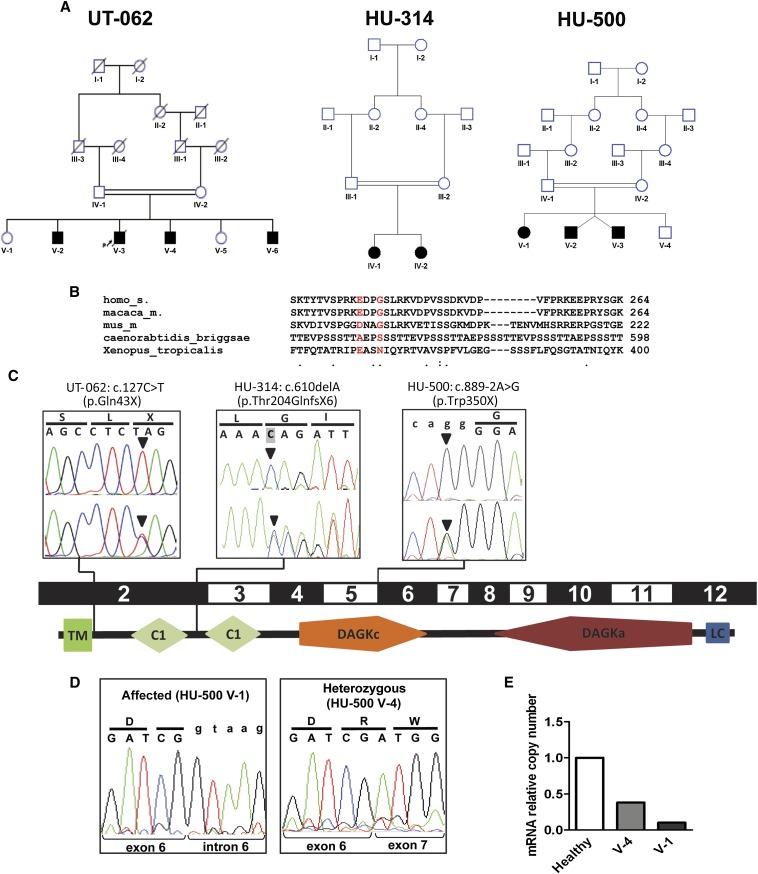
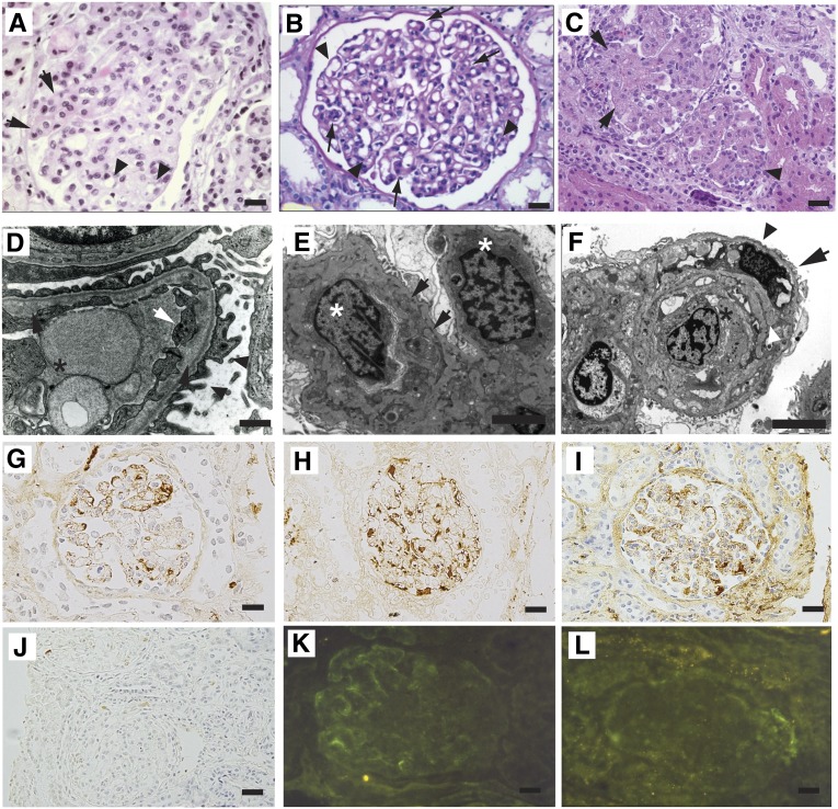
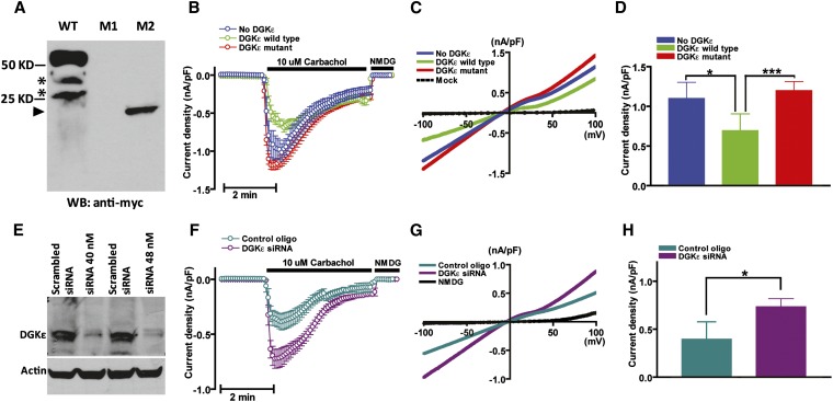
Renal microangiopathies and membranoproliferative GN (MPGN) can manifest similar clinical presentations and histology, suggesting the possibility of a common underlying mechanism in some cases. Here, we performed homozygosity mapping and whole exome sequencing in a Turkish consanguineous family and identified DGKE gene variants as the cause of a membranoproliferative-like glomerular microangiopathy. Furthermore, we identified two additional DGKE variants in a cohort of 142 unrelated patients diagnosed with membranoproliferative GN. This gene encodes the diacylglycerol kinase DGKε, which is an intracellular lipid kinase that phosphorylates diacylglycerol to phosphatidic acid. Immunofluorescence confocal microscopy demonstrated that mouse and rat Dgkε colocalizes with the podocyte marker WT1 but not with the endothelial marker CD31. Patch-clamp experiments in human embryonic kidney (HEK293) cells showed that DGKε variants affect the intracellular concentration of diacylglycerol. Taken together, these results not only identify a genetic cause of a glomerular microangiopathy but also suggest that the phosphatidylinositol cycle, which requires DGKE, is critical to the normal function of podocytes.


Figures

Figure 1.
Figure 1.
Representation of the pedigrees described in this study and of the DGKE mutations in relation to the gene exon structure and protein domains. (A) Pedigrees of families UT-062, HU-314, and HU-500. Squares represent males, circles represent females. Black filled symbols indicate the affected status. Double-horizontal bars indicate consanguinity. (B) Sequence alignment across different species of the GPRIN1 gene in correspondence of the two sequence variants detected by exome sequencing in the affected individuals of family UT-062. The reference amino acids are represented in red. Dots on the bottom line represent medium (:) and low (.) evolutionary conservation. The absence of a dot indicates absence of evolutionary conservation. Numbers indicate the position of the last represented amino acid in the corresponding protein. (C) Chromatograms of the three different mutations in the gene DGKE (arrowheads) in the representative affected individuals from the consanguineous pedigrees UT-062, HU-314, and HU-500, in relation to the corresponding coding exons and schematic representation of human DGKε protein structure (according to SMART, http://smart.embl-heidelberg.de). Pedigree identifiers and mutations are reported above the boxes. Upper chromatograms report the homozygous mutations in one affected sibling per each pedigree. Lower chromatograms show the heterozygous mutations in one of the parents from each family. Codons and the corresponding translated amino acids are displayed above the chromatograms. Lowercase letters indicate the intronic sequence. Shading of the first base of the second codon in HU-314 chromatogram indicates the deleted base. (D) Chromatograms of the RT-PCR on mRNA obtained from peripheral leukocytes of one affected individual (V-1) and a heterozygous sibling (V-4) of family HU-500. Intron 6 is retained in the presence of homozygous mutation (left panel), resulting in the transcription of the intron (lowercase letters). Note that in the heterozygous sibling (right panel), only the wild-type allele is detectable, indicating that the amount of mutated mRNA is very low compared with the wild type. Codons and the corresponding translated amino acids are displayed above the chromatograms. Lowercase letters indicate the intronic sequence. (E) Relative quantitation by real-time PCR of the number of DGKE transcripts in leukocytes from an healthy individual, an heterozygous carrier, and the homozygous affected individual HU-500 V-1 showing that the mutated transcript undergoes non-sense–mediated decay. TM, transmembrane domain; C1, protein kinase C conserved region (C1 domain); DAGKc, diacylglycerol kinase catalytic domain; DAGKa, diacylglycerol kinase accessory domain; LC, low complexity region.
Figure 2.
Figure 2.
Loss-of-function mutations in DGKE cause MPGN-like glomerular microangiopathy. (A) Kidney biopsy of patient UT-062 V-6. The represented glomerulus is hypertrophic and hypercellular. Focal capillary obliteration and thickening of the basement membrane can also be noticed (arrows) compared with patent capillaries (arrowheads). Hematoxylin and eosin. (B) Periodic acid–Schiff staining of a specimen from patient HU-314 IV-2 shows focal duplication of the glomerular basement membrane (arrowheads), causing thickening of the basement membranes in a hypertrophic glomerulus. (C) Image of the kidney biopsy from patient HU-500 V-1. The represented glomeruli are hypertrophic, hyperlobulated, and hypercellular (arrowhead) and present obliteration of the vascular spaces with endothelial cell swelling (arrows). Hematoxylin and eosin. (D) Transmission electron microscopy image of a glomerulus of patient UT-062 V-6. The capillary lumen is obliterated by the body of an endothelial cell (asterisk). Endothelial cytoplasmic rim is swollen (white arrow). Lamina rara interna is irregularly widened with flocculent material (black arrows). Foot processes on the epithelial side are partially effaced (arrowheads). (E) Electron micrograph of kidney biopsy of patient HU-500 V-1. Two swollen endothelial cells (asterisks) occlude a capillary lumen. The basement membrane is split (arrows) by the interposition of a mesangial cell. Uranyl acetate and lead citrate. (F) Electron micrograph of the kidney biopsy of patient HU-500 V-2. A swollen endothelial cell (asterisk) obstructs the lumen of a capillary. The basement membrane is split (white arrowhead) by the interposition of a mesangial cell (arrow). The podocyte foot processes are effaced (black arrowhead). Uranyl acetate and lead citrate. (G–J) Immunoperoxidase staining of biopsies from patient HU-500 V-1. Segmental deposition of IgM (G) and patchy deposition of IgG (H) are visible. Less intense stain was obtained with anti-C1q (I) and no C3 deposition was detected (J). (K and L) Immunofluorescence microscopy of biopsy from patient UT-062 V-6 showing week peripheral segmental deposition of IgM (K) and no intraglomerular deposits of C3 (L). Scale bar, 20 µm in A–C; 1 µm in E and F; 20 µm in G–L.
Figure 3.
Figure 3.
DGKε regulates intracellular DAG levels. (A) Western blot of HEK293T cells transfected with expression vector of myc-tagged wild-type human DGKε (WT) and c.127C>T (M1) and c.610delA (M2) mutants. Nonspecific bands (asterisks) are observed in the WT lane. No peptide was observed overexpressing the c.127C>T mutant. A truncated protein of the expected size (arrowhead at approximately 23 kD) was produced when cells were transfected with the c.610delA mutant. (B) Time course of carbachol (CCh, 10 μM)-induced whole-cell inward TRPC6 current density (nA/pF at −100 mV, mean ± SD, n=20) measured in HEK293T cells transfected with empty expression vector (blue trace), wild-type (green), and c.610delA mutant (red) human DGKε. (C) Current-voltage relationships of currents from B. (D) Mean ± SD of peak inward current density from B. (E) Western blot of HEK293 cells transfected with 40 or 80 nmol of a pool of DGKE-targeting siRNAs or a nontargeting siRNA as control. (F) Time course of carbachol–induced (CCh, 10 μM) whole-cell inward TRPC6 current density measured in HEK293T cells transfected with targeting siRNA (40 nmol, dark green) or nontargeting siRNA as a control (purple). (G) Current-voltage relationships of currents from F. Mean ± SD (n=20) of peak inward current density from F. *P<0.05; ***P<0.001.

Comment in

References

    1. Braun MC, Licht C, Strife CF: Membranoproliferative glomerulonephritis. In: Pediatric Nephrology, edited by Avner ED, Harmon WH, Niaudet P, Yoshikawa N, Berlin, Springer, 2009, pp 783–797
    1. Sethi S, Fervenza FC: Membranoproliferative glomerulonephritis: Pathogenetic heterogeneity and proposal for a new classification. Semin Nephrol 31: 341–348, 2011 - PubMed
    1. Bakkaloglu A, Söylemezoglu O, Tinaztepe K, Saatçi U, Söylemezoglu F: Familial membranoproliferative glomerulonephritis. Nephrol Dial Transplant 10: 21–24, 1995 - PubMed
    1. Berry PL, McEnery PT, McAdams AJ, West CD: Membranoproliferative glomerulonephritis in two sibships. Clin Nephrol 16: 101–106, 1981 - PubMed
    1. Bogdanović RM, Dimitrijević JZ, Nikolić VN, Ognjanović MV, Rodić BD, Slavković BV: Membranoproliferative glomerulonephritis in two siblings: Report and literature review. Pediatr Nephrol 14: 400–405, 2000 - PubMed

Publication types

MeSH terms