. 2013 Feb;24(3):385-92.
doi: 10.1681/ASN.2012101031. Epub 2012 Dec 28.

TGF-β1-containing exosomes from injured epithelial cells activate fibroblasts to initiate tissue regenerative responses and fibrosis

Affiliations

TGF-β1-containing exosomes from injured epithelial cells activate fibroblasts to initiate tissue regenerative responses and fibrosis

Fernanda T Borges et al. J Am Soc Nephrol. 2013 Feb.

Abstract

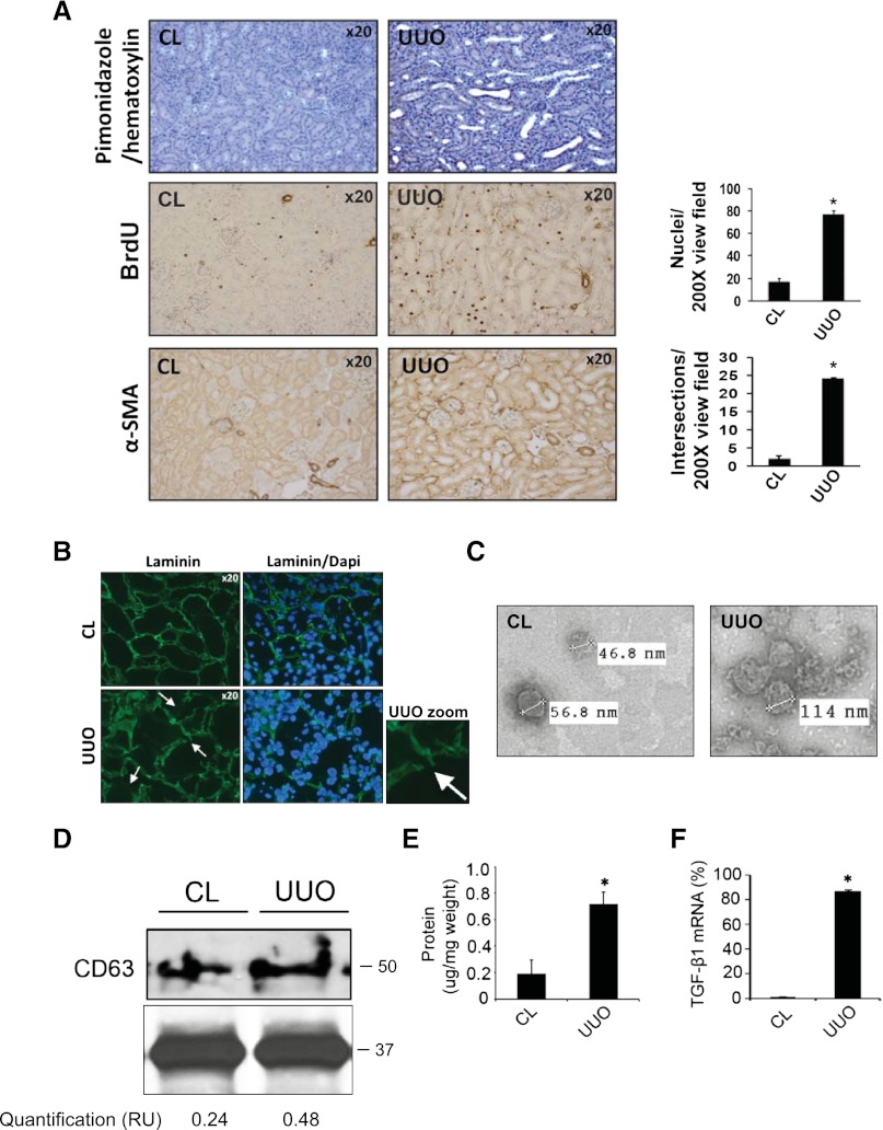
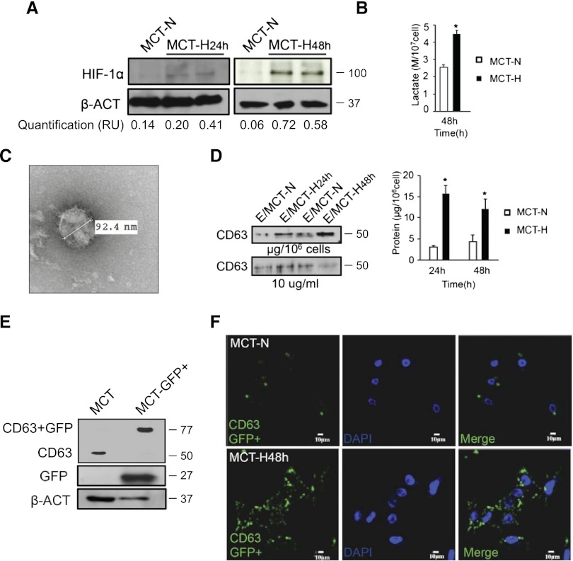
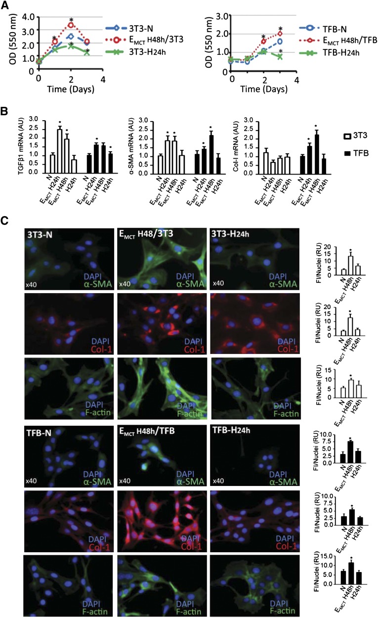
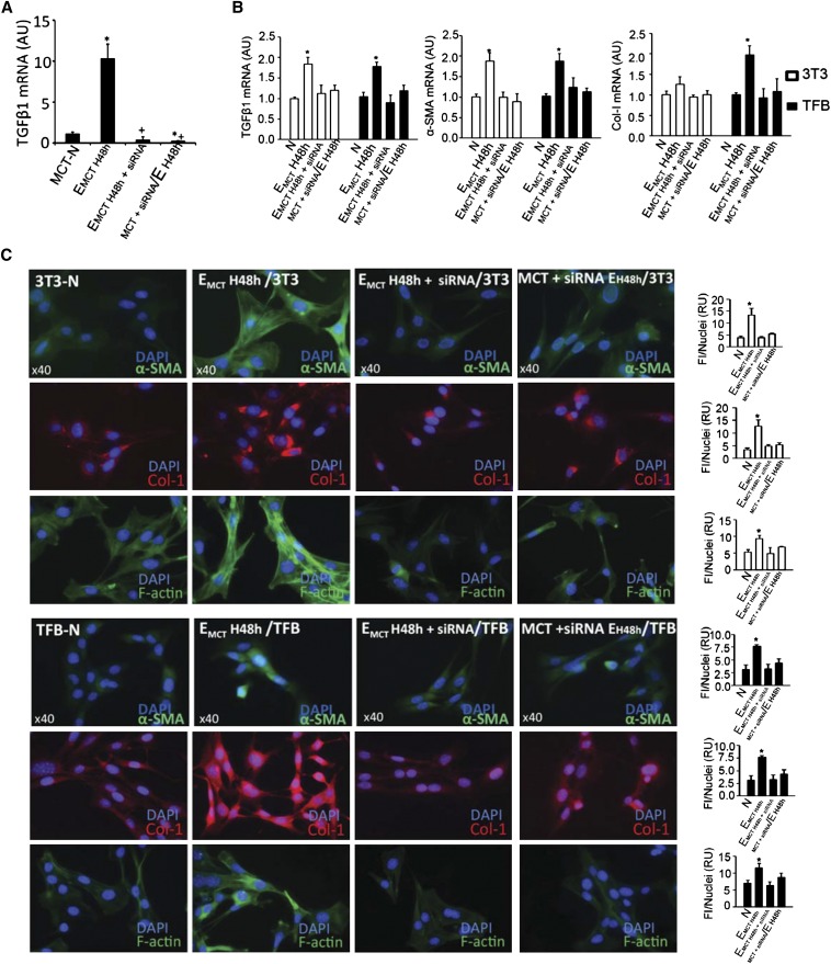
Hypoxia is associated with tissue injury and fibrosis but its functional role in fibroblast activation and tissue repair/regeneration is unknown. Using kidney injury as a model system, we demonstrate that injured epithelial cells produce an increased number of exosomes with defined genetic information to activate fibroblasts. Exosomes released by injured epithelial cells promote proliferation, α-smooth muscle actin expression, F-actin expression, and type I collagen production in fibroblasts. Fibroblast activation is dependent on exosomes delivering TGF-β1 mRNA among other yet to be identified moieties. This study suggests that TGF-β1 mRNA transported by exosomes constitutes a rapid response to initiate tissue repair/regenerative responses and activation of fibroblasts when resident parenchyma is injured. The results also inform potential utility of exosome-targeted therapies to control tissue fibrosis.


Figures

Figure 1.
Figure 1.
An increased number of exosomes are released by fibrotic kidneys that contain TGF-β1 mRNA. (A) Hypoxyprobe, bromodeoxyuridine, and α-SMA staining in kidney cortex after 2 days of UUO surgery using the contralateral kidney as control. Graphs on the right represent the respective quantifications using the number of positive intersections per field or the number of positive nuclei. (B) Immunofluorescence staining with laminin antibody at UUO day 2. Arrows point to the site of tubular basement membrane disruption. (C) Morphologic characterization using electronic microscopy of exosomes extracted from whole contralateral or UUO kidneys of operated mice. The size of exosomes is quantified and expressed in nanometers. (D) Protein blot of exosomes extracted from contralateral and UUO kidneys of operated mice using CD63 antibody and β-actin as loading control with densitometry quantification. Protein extracts prepared from exosomes are normalized by protein extract volume/tissue weight. (E) Protein extracts prepared from exosomes are normalized by protein extract volume/tissue weight and expressed in micrograms per milligram of weight. (F) TGF-β1 mRNA content in exosomes extracted from mouse kidneys after 2 days of UUO. Results are depicted in percentages of TGF-β1 mRNA expression in UUO compared with CL kidney. CL, kidney from contralateral side. UUO, kidney after UUO surgery. Original magnification, ×200 in A. *P<0.05.
Figure 2.
Figure 2.
Hypoxia induces increased production of exosomes by TECs. (A) HIF-1α Western blot in MCT cellular extracts cultured under normoxic or hypoxic conditions. The bands are quantified and the HIF-1α and β-actin ratio is expressed in relative units (RUs). (B) Lactate quantification in culture medium from cells cultured under normoxic (N) and hypoxic (H) conditions. (C) Morphologic characterization of MCT exosomes by electronic microscopy. The size of exosomes is measured and expressed in nanometers. (D) Total exosome protein quantification (right graph). Protein extracts prepared from exosomes are normalized by protein extract volume per cell number and expressed in micrograms per 106 cells. CD63 Western blot analysis shows increased CD63 expression in hypoxia (left panel). Protein extracts prepared from exosomes are normalized by protein extract volume per cell culture number (micrograms per 106 cells; upper panel) or by concentration (10 µg; lower panel) and are assessed using antibody against exosomal protein marker CD63. (E) Western blot using CD63 and GFP antibodies of protein extract from MCT CD63-GFP cells using β-actin expression as loading control. (F) Confocal microscopy for GFP presence in MCT CD63-GFP cells cultured under normoxic or hypoxic conditions. Green signal represents the fusion protein CD63-GFP.
Figure 3.
Figure 3.
Hypoxic TEC-derived exosomes lead to fibroblast activation. (A) MTT assay shows proliferation of 3T3 and TFB fibroblasts in normoxia (N), hypoxia (H24h), or exposed to exosomes extracted from MCT cells cultured under hypoxia for 48 hours (EMCT H48h). (B) TGF-β1, α-SMA, and collagen-1 expression in normoxic (N) 3T3 and TFB fibroblasts that are treated with hypoxic exosomes secreted from MCT cells after 24 hours (EMCT H24h) and 48 hours (EMCT H48h) of hypoxia. Results are expressed in arbitrary units (AUs). (C) Immunofluorescence of α-SMA, collagen-1 (Col-1), and F-actin in 3T3 and TFB fibroblasts in normoxia (N), hypoxia (H24h), or treated with exosomes from MCT hypoxic cells (EMCT H48h). Fluorescence intensity per nuclei (FI/Nuclei) is quantified and expressed in relative units (RUs). *Significantly different from normoxic control cells (P<0.05).
Figure 4.
Figure 4.
Hypoxic TEC exosome-derived TGF-β1 mRNA is functionally important for the activation of fibroblasts. (A) TGF-β1 expression in MCT cells under different experimental conditions. In EMCT/H48h, MCT cells are exposed to hypoxia for 48 hours, the culture medium is collected, and exosomes are obtained for RNA extraction. In EMCT 48h + siRNA, MCT cells are exposed to hypoxia for 48 hours, and exosomes are extracted and treated with TGF-β1 siRNA. In MCT+ siRNA/EH48h, MCT cells are treated with siRNA, exposed to hypoxia for 48 hours, the culture medium is collected, and exosomes are obtained for RNA extraction; after this period, RNA is extracted for TGF-β1 expression analyses. *P<0.05 compared with MCT cells under normoxia (MCT-N); +P<0.05 compared with EMCT H48h. (B) TGF-β1, α-SMA, and collagen-1 expression and (C) immunofluorescence for α-SMA, collagen-1 (Col1a1), and F-actin in 3T3 and TFB normoxic (N) fibroblasts treated with hypoxic exosomes extracted from MCT cells under 48 hours of hypoxia (EMCT H48h). Exosomes were extracted from MCT cells cultured for 48 h under hypoxia and were then electroporated with TGF-β1 siRNA (EMCT H48h + siRNA), and exosomes extracted from MCT cells treated with TGF-β1 siRNA for 48 hours of hypoxia (MCT + siRNA/EH48h). Results are expressed in arbitrary units (AUs) for gene expression and the fluorescence intensity per nuclei (FI/Nuclei) is quantified and expressed in relative units (RUs) for immunofluorescence. *P<0.05.

Comment in

References

    1. Lee SB, Kalluri R: Mechanistic connection between inflammation and fibrosis. Kidney Int Suppl 119: S22–S26, 2010 - PMC - PubMed
    1. Tomasek JJ, Gabbiani G, Hinz B, Chaponnier C, Brown RA: Myofibroblasts and mechano-regulation of connective tissue remodelling. Nat Rev Mol Cell Biol 3: 349–363, 2002 - PubMed
    1. Okada H, Kalluri R: Cellular and molecular pathways that lead to progression and regression of renal fibrogenesis. Curr Mol Med 5: 467–474, 2005 - PubMed
    1. Sugimoto H, LeBleu VS, Bosukonda D, Keck P, Taduri G, Bechtel W, Okada H, Carlson W, Jr, Bey P, Rusckowski M, Tampe B, Tampe D, Kanasaki K, Zeisberg M, Kalluri R: Activin-like kinase 3 is important for kidney regeneration and reversal of fibrosis. Nat Med 18: 396–404, 2012 - PMC - PubMed
    1. Coresh J, Astor BC, Greene T, Eknoyan G, Levey AS: Prevalence of chronic kidney disease and decreased kidney function in the adult US population: Third National Health and Nutrition Examination Survey. Am J Kidney Dis 41: 1–12, 2003 - PubMed

Publication types

MeSH terms

Substances