

Review
doi: 10.1007/s10557-012-6430-0.
The late Na+ current--origin and pathophysiological relevance
Affiliations
- PMID: 23274937
- PMCID: PMC3555240
- DOI: 10.1007/s10557-012-6430-0
Item in Clipboard
Review
The late Na+ current--origin and pathophysiological relevance
Cardiovasc Drugs Ther.
2013 Feb.
No abstract available
Figures
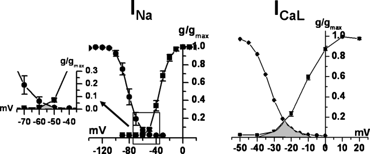
Comparison of the membrane potential “window” for INa and ICaL in rat ventricular myocytes. Steady-state activation and inactivation curves for INa (left) and ICaL (right). The “window”, i.e. the membrane potential range in which activation and inactivation overlap, is filled in grey. INa window (zoomed in the inset) is smaller and at more negative potentials than ICaL one. (from Rocchetti et al. unpublished)
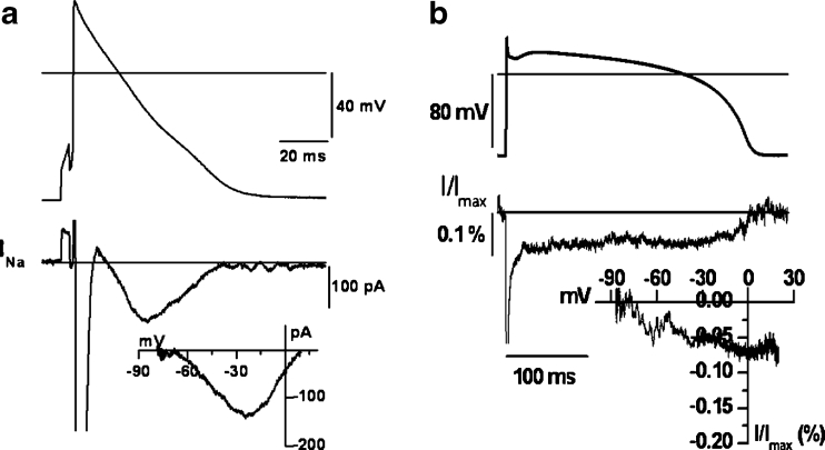
TTX-sensitive current (lower panels, largely representative of INa) elicited by clamping the membrane with the action potential waveform (upper panel) (AP-clamp); the inset in each panel shows the dynamic I/V relation, obtained by plotting current as a function of membrane potential. TTX-sensitive current flowing during repolarization includes all sustained Na+ components. a recording in a rat myocyte, clamped with its own action potential (Rocchetti et al. unpublished); b recording in a cell line (HEK239) transfected with NaV1.5 (α and β subunits) and clamped with a human ventricular action potential (from ref [12], modified). INaT is truncated in all recordings
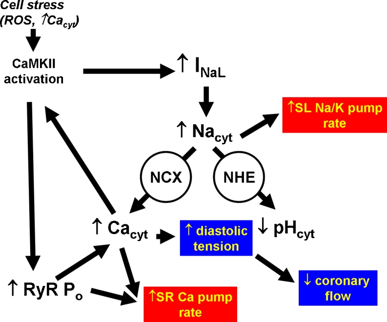
Network of events linking INaL enhancements to its pleiotropic effects. Notably, many events are linked in positive feed back loops, likely to amplify and sustain the network once it has been initiated. Experimental observations (see text) suggest that INaL enhancement and CaMKII activation are key elements in this process. Events in red boxes imply increased ATP consumption, those in blue boxes reduced oxygen supply
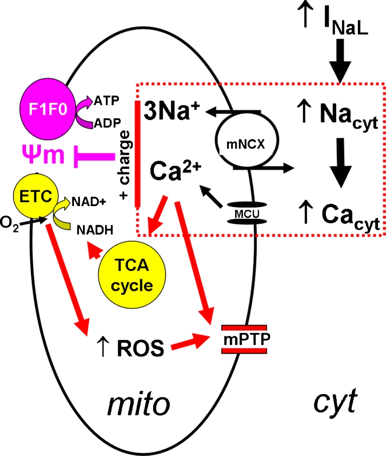
A plausible sequence of events coupling INaL enhancement to mitochondrial damage. Functions primarily depressed upon INaL enhancement are in purple, those upregulated to compensate in yellow. Elevated cytosolic Ca2+ (Cacyt) loads the mitochondria through the Ca2+ uniporter (MCU) and stimulates NADH production by the tricarboxilic acids (TCA) cycle. Excess mitochondrial Ca2+ is partially removed by mitochondrial NCX (mNCX), driven by high cytosolic Na+ [57]. This chain of events (in the dotted box) results in positive charge influx, which tends to dissipate the electrical gradient (Ψm) that drives ATP synthesis by the ATP-synthase (F1F0) [64]. The resulting drop in the ATP/ADP ratio is compensated by increasing the electron transport rate (by ETC), fuelled by NADH [57]. In this scheme NADH and ATP levels may be preserved up to a limit [57], but at the cost of increased substrate consumption and ROS production by ETC [43]. Concomitantly elevated mitochondrial Ca2+ and ROS facilitate opening of mPTP [43], eventually resulting in loss of membrane selectivity, swelling and disruption of the outer mitochondrial membrane. Release of cyrochrome C and other components of the outer membrane space triggers cell apoptosis [64]
References
Publication types
MeSH terms
Substances
LinkOut - more resources
Full Text Sources
Medical
