

Simvastatin protects hepatocytes from apoptosis by suppressing the TNF-α/caspase-3 signaling pathway in mice with burn injury
- PMID: 23275311
- PMCID: PMC4102342
- DOI: 10.1097/SLA.0b013e318273fdca
Simvastatin protects hepatocytes from apoptosis by suppressing the TNF-α/caspase-3 signaling pathway in mice with burn injury
Abstract
Objective: To investigate the liver cellular apoptosis in response to burn injury and find out if statin treatment can ameliorate this process. The hypothesis is that statin may modulate apoptosis-related gene expression and thereby reduce hepatocytic apoptosis after burn injury.
Methods: Mice were subjected to 30% full-thickness burn injury and then treated either with or without simvastatin. The livers were harvested for histological assessment and determinations of gene expression. To investigate the mechanism involved, tumor necrosis factor (TNF)-α and caspase-3 knockout (KO) mice were also used to evaluate the effects of burn injury and simvastatin treatment on burn-induced liver injury. The effects of simvastatin on TNF-α and caspase-3 expressions were also evaluated in cultured mouse hepatocytes.
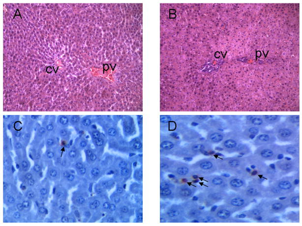
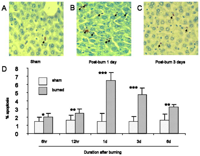
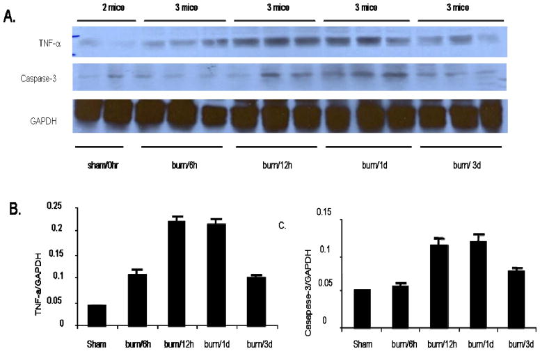
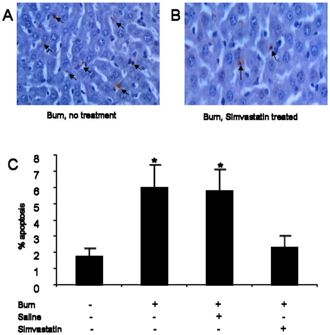
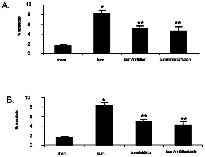
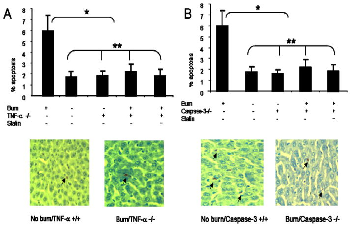
Results: Burn injury induced significant liver damage, which was indicated by striking levels of apoptosis. Simvastatin reduced the apoptotic index in the livers of mice with burn injury and this effect could be abrogated by TNF-α or caspase-3 inhibitors. Simvastatin also decreased burn-induced TNF-α and caspase-3 expression in the liver. TNF-α and caspase-3 KO mice demonstrated lower levels of apoptotic hepatocytes in response to burn, and simvastatin did not further decrease hepatocyte apoptosis in either strain of KO mice. An in vitro study demonstrated that simvastatin suppresses TNF-α and caspase-3 expression in primary cultures of mouse hepatocytes.
Conclusions: Simvastatin reduces mouse hepatocyte apoptosis by suppressing expression of the TNF-α/caspase-3 pathway.
Conflict of interest statement
None.
Figures

Similar articles
-
Simvastatin reduces burn injury-induced splenic apoptosis via downregulation of the TNF-α/NF-κB pathway.Ann Surg. 2015 May;261(5):1006-12. doi: 10.1097/SLA.0000000000000764. Ann Surg. 2015. PMID: 24950285 Free PMC article.
-
Naofen promotes TNF-α-mediated apoptosis of hepatocytes by activating caspase-3 in lipopolysaccharide-treated rats.World J Gastroenterol. 2014 May 7;20(17):4963-71. doi: 10.3748/wjg.v20.i17.4963. World J Gastroenterol. 2014. PMID: 24803807 Free PMC article.
-
AKT activator SC79 protects hepatocytes from TNF-α-mediated apoptosis and alleviates d-Gal/LPS-induced liver injury.Am J Physiol Gastrointest Liver Physiol. 2019 Mar 1;316(3):G387-G396. doi: 10.1152/ajpgi.00350.2018. Epub 2019 Jan 10. Am J Physiol Gastrointest Liver Physiol. 2019. PMID: 30629471
-
Differential caspase-9-dependent signaling pathway between tumor necrosis factor receptor- and Fas-mediated hepatocyte apoptosis in mice.Liver Int. 2006 Feb;26(1):137-46. doi: 10.1111/j.1478-3231.2005.01195.x. Liver Int. 2006. PMID: 16420519
-
Tumor necrosis factor signaling in hepatocyte apoptosis.J Gastroenterol Hepatol. 2007 Jun;22 Suppl 1:S43-4. doi: 10.1111/j.1440-1746.2006.04645.x. J Gastroenterol Hepatol. 2007. PMID: 17567463 Review.
Cited by
-
Advanced age heightens hepatic damage in a murine model of scald burn injury.J Trauma Acute Care Surg. 2021 Apr 1;90(4):731-737. doi: 10.1097/TA.0000000000003048. J Trauma Acute Care Surg. 2021. PMID: 33306599 Free PMC article.
-
Simvastatin Inhibits Endotoxin-Induced Apoptosis in Liver and Spleen Through Up-Regulation of Survivin/NF-κB/p65 Expression.Front Pharmacol. 2019 Feb 15;10:54. doi: 10.3389/fphar.2019.00054. eCollection 2019. Front Pharmacol. 2019. PMID: 30828299 Free PMC article.
-
Evidence for simvastatin anti-inflammatory actions based on quantitative analyses of NETosis and other inflammation/oxidation markers.Results Immunol. 2014 Mar 25;4:14-22. doi: 10.1016/j.rinim.2014.03.001. eCollection 2014. Results Immunol. 2014. PMID: 24809006 Free PMC article.
-
Reducing PKCδ inhibits tumor growth through growth hormone by inhibiting PKA/CREB/ERK signaling pathway in pituitary adenoma.Sci Rep. 2025 Apr 3;15(1):11461. doi: 10.1038/s41598-025-95865-3. Sci Rep. 2025. Retraction in: Sci Rep. 2025 Jul 15;15(1):25508. doi: 10.1038/s41598-025-10487-z. PMID: 40181069 Free PMC article. Retracted.
-
Membrane potential-dependent uptake of 18F-triphenylphosphonium--a new voltage sensor as an imaging agent for detecting burn-induced apoptosis.J Surg Res. 2014 May 15;188(2):473-9. doi: 10.1016/j.jss.2014.01.011. Epub 2014 Jan 11. J Surg Res. 2014. PMID: 24582214 Free PMC article.
References
-
- Jeschke MG, Herndon DN, Ebener C, et al. Nutritional intervention high in vitamins, protein, amino acids, and omega3 fatty acids improves protein metabolism during the hypermetabolic state after thermal injury. Arch Surg. 2001;136(11):1301–6. - PubMed
-
- Sener G, Kabasakal L, Cetinel S, et al. Leukotriene receptor blocker montelukast protects against burn-induced oxidative injury of the skin and remote organs. Burns. 2005;31(5):587–96. - PubMed
-
- Zhou ZB, Ding HQ, Qin FJ, et al. Effect of Zn7-metallothionein on oxidative stress in liver of rats with severe thermal injury. Acta Pharmacol Sin. 2003;24(8):764–70. - PubMed
-
- Sener G, Sehirli AO, Gedik N, et al. Rosiglitazone, a PPAR-gamma ligand, protects against burn-induced oxidative injury of remote organs. Burns. 2007;33(5):587–93. - PubMed
Publication types
MeSH terms
Substances
Grants and funding
LinkOut - more resources
Full Text Sources
Other Literature Sources
Medical
Research Materials
