Review
. 2012 Dec;53(6):518-28.
doi: 10.3325/cmj.2012.53.518.

Astrocytes revisited: concise historic outlook on glutamate homeostasis and signaling

Affiliations
Review

Astrocytes revisited: concise historic outlook on glutamate homeostasis and signaling

Vladimir Parpura et al. Croat Med J. 2012 Dec.

Abstract

Astroglia is a main type of brain neuroglia, which includes many cell sub-types that differ in their morphology and physiological properties and yet are united by the main function, which is the maintenance of brain homeostasis. Astrocytes employ a variety of mechanisms for communicating with neuronal networks. The communication mediated by neurotransmitter glutamate has received a particular attention. Glutamate is de novo synthesized exclusively in astrocytes; astroglia-derived glutamine is the source of glutamate for neurons. Glutamate is released from both neurons and astroglia through exocytosis, although various other mechanisms may also play a role. Glutamate-activated specific receptors trigger excitatory responses in neurons and astroglia. Here we overview main properties of glutamatergic transmission in neuronal-glial networks and identify some future challenges facing the field.


Figures
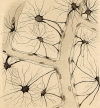
Figure 1
Figure 1
Neuroglial cells drawn by Camillo Golgi. Cells were stained using the silver-chromate technique. Individual star-shaped astrocytes form astroglial network and numerous contacts (the endfeet) with brain capillaries. The image [reproduced from (6)] was kindly provided by Prof. Paolo Mozzarello.
Figure 2
Figure 2
Evolution of neuroglia.
Figure 3
Figure 3
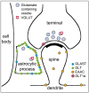
Schematic representation of the localization of glutamate transporters at the hippocampal tripartite synapse. Glutamatergic synapses in the hippocampus are contacted by astrocytes. GLAST and GLT are present in the astrocytic membranes at high density, especially toward the neuropil. Presynaptic terminals specifically contain GLT1a variant. Quantitative information on EAAC is not available. Neurons and astrocytes possess vesicular glutamate transporter (VGLUT)-laden vesicles which contain glutamate. Modified from (18).
Figure 4
Figure 4
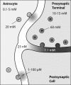
Glutamate concentration in different cellular and extracellular compartments at the tripartite synapse. Modified from (29).
Figure 5
Figure 5
High frequency (50 Hz) electrical stimulation of mossy fibers evokes [Ca2+]i responses in CA3 region astrocytes. (A) Drawing shows positioning of stimulating electrode (Stim) and imaged area (rectangular area) shown in B-F with respect to microanatomy of transverse hippocampal slice. Stimulating electrode is placed in dentate gyrus (DG) to depolarize axons of granule cells (g), which synapse onto pyramidal neurons (p) in CA3 region. Cell bodies of pyramidal neurons form palisades (dotted outlines) and are surrounded by astrocytes (a). (B) Resting fluorescence observed within CA3 region after loading slices with Ca2+ indicator fluo-3 (32 frame average). Note the stratum pyramidale (palisades) formed by neuronal somata (left 2/3 of the image), while stratum lucidum lies to the far right. The squares numbered 1-5 indicate astrocytes, while 6 indicates a neuron.(C) GFAP immunofluorescent reactivity of the field shown in B acquired after the Ca2+ imaging sequence shown in D-F (single frames). Arrows and arrowhead indicate astrocytic somata and processes, respectively. (D) The earliest response in CA3 region following electrical stimulation of dentate gyrus. The arrows indicate the active horizontal scan line at the time of stimulus onset (t = 0 seconds). Thus, the portion of the image above the line represents fluorescence before stimulation, while the portion of the image below the line reports on fluorescence intensities after electrical stimulation, showing [Ca2+]i increases in pyramidal cell bodies and fine neuronal processes. (E) After an additional 2 seconds of stimulation (t = 4 seconds), the pyramidal cell bodies exhibited large [Ca2+]i increases, but also many GFAP-positive cell bodies and processes showed [Ca2+]i increases. Arrows and arrowhead in E and F correspond to those in C. (F) After 4 seconds of stimulation (t = 6 seconds) almost all astrocytes within the imaging area responded with [Ca2+]i increases. Scale bar, 20 μm. Modified from (46). Data courtesy of Dr Stephen J. Smith, Stanford University.
Figure 6
Figure 6
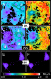
Bradykinin causes a glutamate-mediated accumulation of internal Ca 2+ in neurons co-cultured with astrocytes. The [Ca2+]i in neocortical neurons (dotted circles in A and C) and astrocytes (a in A and C) was monitored using fura-2. (A) Mixed culture at rest. (B) Application of bradykinin (BK, 1 μM, 75 seconds) caused an elevation in [Ca2+]i in astrocytes and neurons. (C) However, when co-cultures were bathed in presence of the broad spectrum GluR antagonist D-glutamylglycine (DGG), application of bradykinin did not significantly alter neuronal [Ca2+]i calcium levels, even though bradykinin elevated the astrocytic [Ca2+]i (D). (E-F) Bradykinin failed to elevate [Ca2+]i in neurons devoid of surrounding astrocytes. Color scale indicates pseudocolor representation of [Ca2+]i, by fura-2 emission ratio ranging from 0 to 2.0. Modified from (55).

Similar articles

Cited by

References

    1. Virchow R. Cellular pathology: as based upon physiological and pathological histology. Twenty lectures delivered in the pathological institute of Berlin during the months of February, March and April, 1858 [in German]. First edition ed. Berlin: August Hirschwald; 1858.
    1. Kettenmann H, Verkhratsky A. Neuroglia: the 150 years after. Trends Neurosci. 2008;31:653–9. doi: 10.1016/j.tins.2008.09.003. - DOI - PubMed
    1. Verkhratsky A. Patching the glia reveals the functional organisation of the brain. Pflugers Arch. 2006;453:411–20. doi: 10.1007/s00424-006-0099-9. - DOI - PubMed
    1. Verkhratsky A, Parpura V, Rodriguez JJ. Where the thoughts dwell: the physiology of neuronal-glial “diffuse neural net”. Brain Res Rev. 2011;66:133–51. doi: 10.1016/j.brainresrev.2010.05.002. - DOI - PubMed
    1. Golgi C. On structure of the gray matter of the brain. Gazzetta Medica Italiana Lombardia. 1873;33:244–6. [in Italian]

Publication types