. 2013 Feb 8;288(6):3977-88.
doi: 10.1074/jbc.M112.436311. Epub 2012 Dec 30.

Yes-associated protein isoform 1 (Yap1) promotes cardiomyocyte survival and growth to protect against myocardial ischemic injury

Affiliations

Yes-associated protein isoform 1 (Yap1) promotes cardiomyocyte survival and growth to protect against myocardial ischemic injury

Dominic P Del Re et al. J Biol Chem. .

Abstract

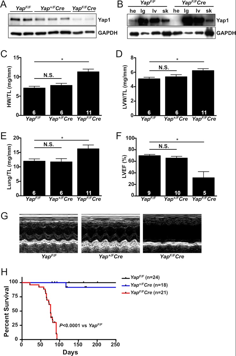
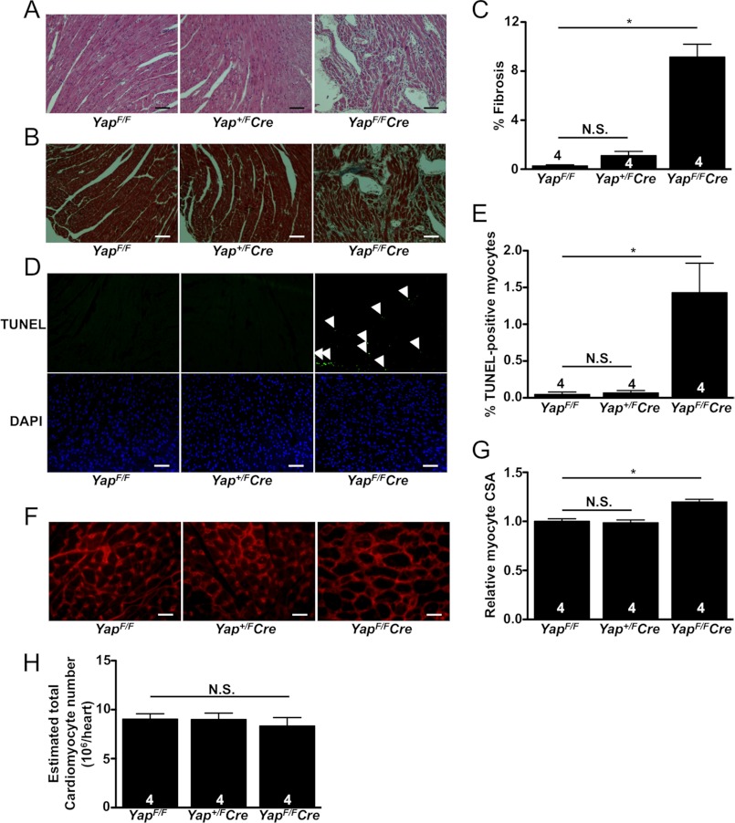
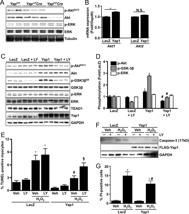
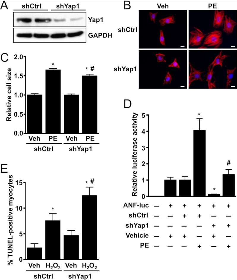
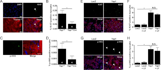
Yap1 is an important regulator of cardiomyocyte proliferation and embryonic heart development, yet the function of endogenous Yap1 in the adult heart remains unknown. We studied the role of Yap1 in maintaining basal cardiac function and in modulating injury after chronic myocardial infarction (MI). Cardiomyocyte-specific homozygous inactivation of Yap1 in the postnatal heart (Yap(F/F)Cre) elicited increased myocyte apoptosis and fibrosis, dilated cardiomyopathy, and premature death. Heterozygous deletion (Yap(+/F)Cre) did not cause an overt cardiac phenotype compared with Yap(F/F) control mice at base line. In response to stress (MI), nuclear Yap1 was found selectively in the border zone and not in the remote area of the heart. After chronic MI (28 days), Yap(+/F)Cre mice had significantly increased myocyte apoptosis and fibrosis, with attenuated compensatory cardiomyocyte hypertrophy, and further impaired function versus Yap(+/F) control mice. Studies in isolated cardiomyocytes demonstrated that Yap1 expression is sufficient to promote increased cell size and hypertrophic gene expression and protected cardiomyocytes against H(2)O(2)-induced cell death, whereas Yap1 depletion attenuated phenylephrine-induced hypertrophy and augmented apoptosis. Finally, we observed a significant decrease in cardiomyocyte proliferation in Yap(+/F)Cre hearts compared with Yap(+/F) controls after MI and demonstrated that Yap1 is sufficient to promote cardiomyocyte proliferation in isolated cardiomyocytes. Our findings suggest that Yap1 is critical for basal heart homeostasis and that Yap1 deficiency exacerbates injury in response to chronic MI.


Figures

FIGURE 1.
FIGURE 1.
Base-line cardiac characterization of Yap1-deficient mice. A, shown is immunoblot analysis of Yap1 expression in ventricular tissue of YapF/F, Yap+/FCre, and YapF/FCre adult (8-week-old) mice. B, shown is immunoblot analysis of Yap1 expression in the heart (he), lung (lg), liver (lv), and skeletal muscle (sk) from 8-week-old YapF/F control and YapF/FCre mice. C—E, heart weight (HW), left ventricular weight (LVW), and lung weight were measured and normalized to tibia length (TL) in adult mice. F, echocardiography was performed, and LVEF was determined. Values are the means ± S.E. *, p < 0.05. N.S. = not significant. G, representative M-mode tracings are shown. H, shown is a Kaplan-Meier survival curve demonstrating the premature death of YapF/FCre mice. Log-rank (Mantel-Cox) test for survival, YapF/FCre versus YapF/F, p < 0.0001.
FIGURE 2.
FIGURE 2.
Histological analysis of Yap1-deficient mice. A–G, ventricles from adult mice were fixed, sectioned, and stained. A, shown is hematoxylin and eosin staining demonstrating disorganized cardiomyocytes and increased cell infiltration in YapF/FCre hearts. B and C, Masson's Trichrome staining revealed a significant increase in interstitial fibrosis in YapF/FCre hearts. D and E, TUNEL shows a significant increase in apoptotic cardiomyocytes in YapF/FCre hearts. Arrows indicate TUNEL-positive nuclei. F and G, wheat germ agglutinin (WGA) staining reveals increased myocyte CSA in YapF/FCre hearts. H, shown is the estimated number of cardiomyocytes present in the hearts of Yap1 mutant and control mice. Values are the means ± S.E. *, p < 0.05. N.S. = not significant. Scale bars, 100 μm.
FIGURE 3.
FIGURE 3.
Yap1 promotes cardiomyocyte survival through up-regulation of Akt. A, representative immunoblots detect p-Akt(S473), total Akt, p-ERK(T202/Y204), and total ERK from Yap+/F, Yap+/FCre, and YapF/FCre hearts. B, neonatal rat cardiomyocytes were transduced with LacZ control or Yap1 adenovirus, and quantitative real-time-PCR was performed to determine mRNA expression of Akt1 and Akt2. Results are normalized to Gapdh expression. *, p < 0.05. N.S. = not significant. C, cardiomyocytes were transduced with LacZ or Yap1 adenovirus and treated with LY294002 (10 μm) or vehicle. Representative immunoblots are shown. D, quantification of results was obtained from C. *, p < 0.05 versus LacZ. #, p < 0.05 versus Yap1. E, cardiomyocytes were transduced with LacZ or Yap1 adenovirus and treated with LY294002 (10 μm) or vehicle. Cells were then treated with vehicle or H2O2 (100 μmol/liter), and apoptosis was evaluated by TUNEL. Values are the means ± S.E. *, p < 0.05 versus LacZ + vehicle. #, p < 0.05 versus LacZ + H2O2 + vehicle. §, p < 0.05 versus Yap1 + H2O2 + vehicle. F, cardiomyocyte apoptosis was also determined by detection of cleaved caspase-3 (17kD). A representative immunoblot is shown. G, cardiomyocyte viability was determined by propidium iodide (PI) uptake after H2O2 (100 μmol/liter) treatment. Values are the means ± S.E. *, p < 0.05 versus LacZ + vehicle. #, p < 0.05 versus LacZ + H2O2.
FIGURE 4.
FIGURE 4.
Yap1-induced cardiomyocyte hypertrophy is Akt-independent. A, cardiomyocytes were transduced with LacZ or Yap1 adenovirus and treated with LY294002 (10 μm), PD98059 (10 μm), or vehicle and stained to detect troponin T. Scale bars, 10 μm. B, quantification of cardiomyocyte surface area is shown. Values are the means ± S.E. *, p < 0.05. N.S. = not significant. C, cardiomyocytes were transduced with LacZ or Yap1 adenovirus, and quantitative real-time-PCR was performed to detect ANF (Nppa), brain natriuretic protein (Nppb), and β-MHC (Myh7) gene expression. Results were normalized to Gapdh. *, p < 0.05. D, cardiomyocytes were transfected with LacZ or Yap1 in combination with ANF-luc reporter plasmid. Cells were treated with LY294002 (10 μm), Akt inhibitor V (triciribine; 10 μm), PD98059 (10 μm), or vehicle control, and luciferase activity was determined. Values are the means ± S.E. *, p < 0.05 versus LacZ + vehicle. N.S. = not significant.
FIGURE 5.
FIGURE 5.
Depletion of Yap1 attenuates cardiomyocyte hypertrophy and promotes apoptosis. A, shown is a representative immunoblot demonstrating endogenous Yap1 knockdown using adenoviral shRNA. Cardiomyocytes were infected with scrambled control (shCtrl) or Yap1-targeted (shYap1) virus, and cells were harvested 72 h later. B, cardiomyocytes were transduced with shCtrl or shYap1, treated with phenylephrine (PE; 100 μm) or vehicle, and stained to detect troponin T. Scale bars, 10 μm. C, quantification of the cardiomyocyte surface area is shown. Values are the means ± S.E. *, p < 0.05 versus shCtrl + vehicle. #, p < 0.05 versus shCtrl + phenylephrine. D, cardiomyocytes were transfected with shCtrl or shYap1 in combination with ANF-luc reporter plasmid. Cells were treated with phenylephrine (100 μm) or vehicle control, and luciferase activity was determined. Values are the means ± S.E. *, p < 0.05 versus shCtrl + vehicle. #, p < 0.05 versus shCtrl + phenylephrine. E, cardiomyocytes were transduced with shCtrl or shYap1 adenovirus and treated with vehicle or H2O2 (100 μmol/liter), and apoptosis was evaluated by TUNEL. Values are the means ± S.E. *, p < 0.05 versus shCtrl + vehicle. #, p < 0.05 versus shCtrl + H2O2.
FIGURE 6.
FIGURE 6.
Yap1 deficiency exacerbates injury after chronic MI. A, shown is a representative immunoblot demonstrating increased Yap1 phosphorylation at Ser-127 and Lats2 at Ser-872 in hearts of 28-day post-MI mice compared with sham-operated controls. B, shown is quantitation of immunoblot results. *, p < 0.05. C and D, nuclear Yap1 is increased in cardiomyocytes present in the border zone but not in the ischemic or remote zones of MI hearts. C, mice were subjected to permanent coronary ligation. Yap1 staining of the ischemic, border, and remote zones after 4 days is shown. Note that Yap1 accumulates in the nucleus in the border zone (B, arrow), but it is excluded from the nucleus in the ischemic (I, arrowhead) and remote zones comparable to control (sham, thin arrow). Green, anti-Yap1 staining; red, anti-troponin T staining; blue, DAPI. Scale bars, 100 μm. D, shown is quantification of nuclear Yap1-positive cardiomyocytes in the border zone. E, heart weight (HW) normalized to tibia length (TL) was determined after 28 days MI. F, heart sections were stained with wheat germ agglutinin, and CSA was determined in the remote region after MI. G, shown are representative images of Masson's Trichrome-stained Yap+/F and Yap+/FCre hearts after 28 days MI. Scale bars, 1 mm. H, shown is quantification of infarct size as a percentage of total endocardial circumference. I, quantification of fibrosis is shown. J, heart sections were stained by TUNEL to detect cardiomyocytes undergoing apoptosis. K, a representative immunoblot demonstrates increased Akt phosphorylation in hearts from Yap+/F but not Yap+/FCre mice subjected to 2-day MI compared with sham operated controls. Values are the means ± S.E. *, p < 0.05. N.S. = not significant.
FIGURE 7.
FIGURE 7.
Cardiac function is further impaired in Yap-deficient hearts after MI. A–F, serial echocardiography was performed at 0, 7, 14, and 28 days post-MI. LVEF (A), fractional shortening (% FS; B), left ventricular end diastolic dimension (LVEDD; C), left ventricular end systolic dimension (LVESD; D), systolic posterior wall thickness (SPWT; E), and diastolic posterior wall thickness (DPWT; F) were determined in Yap+/F and Yap+/FCre mice. Values are the means ± S.E. *, p < 0.05 versus Yap+/F control at the same time point.
FIGURE 8.
FIGURE 8.
Cardiomyocyte proliferation is attenuated in Yap1-deficient hearts after MI. A, heart sections were stained with anti-Ki-67 (green), anti-troponin T (red), and DAPI (blue). The arrow indicates Ki-67-positive myocyte. B, shown is quantification of Ki-67-positive cardiomyocytes detected in Yap+/F and Yap+/FCre hearts. C and D, hearts were stained with anti-phospho(Ser-10)-histone H3 (green), anti-troponin T (red), and DAPI (blue). C, shown is a representative image of p-HH3-positive cardiomyocyte. D, quantification of p-HH3-positive cardiomyocytes is shown. Note that Ki-67 staining generally showed greater sensitivity than p-HH3 staining in our hands. Scale bars, 50 μm. E, neonatal rat cardiomyocytes were treated with Yap1 or LacZ adenovirus and vehicle or LY294002 (10 μm) then stained with anti-Ki-67 (green), anti-troponin T (red), and DAPI (blue). Scale bars, 30 μm. F, quantification of Ki-67-positive myocytes (%). G, cardiomyocytes were transduced with LacZ or Yap1 adenovirus and vehicle or LY294002 (10 μm) then stained with anti-p-HH3 (green), anti-troponin T (red), and DAPI (blue). Arrows indicate p-HH3-positive myocytes. Scale bars, 30 μm. H, quantification of p-HH3-positive cardiomyocytes (%) is shown. Values are the means ± S.E. *, p < 0.05. N.S. = not significant.

Similar articles

Cited by

References

    1. Velagaleti R. S., Pencina M. J., Murabito J. M., Wang T. J., Parikh N. I., D'Agostino R. B., Levy D., Kannel W. B., Vasan R. S. (2008) Long term trends in the incidence of heart failure after myocardial infarction. Circulation 118, 2057–2062 - PMC - PubMed
    1. Narula J., Haider N., Virmani R., DiSalvo T. G., Kolodgie F. D., Hajjar R. J., Schmidt U., Semigran M. J., Dec G. W., Khaw B. A. (1996) Apoptosis in myocytes in end-stage heart failure. N. Engl. J. Med. 335, 1182–1189 - PubMed
    1. Olivetti G., Abbi R., Quaini F., Kajstura J., Cheng W., Nitahara J. A., Quaini E., Di Loreto C., Beltrami C. A., Krajewski S., Reed J. C., Anversa P. (1997) Apoptosis in the failing human heart. N. Engl. J. Med. 336, 1131–1141 - PubMed
    1. Mani K., Kitsis R. N. (2003) Myocyte apoptosis. Programming ventricular remodeling. J. Am. Coll. Cardiol. 41, 761–764 - PubMed
    1. Pan D. (2010) The Hippo signaling pathway in development and cancer. Dev. Cell 19, 491–505 - PMC - PubMed

Publication types

MeSH terms