Review
. 2013 Jan;193(1):1-50.
doi: 10.1534/genetics.112.143362.

Lipid droplets and peroxisomes: key players in cellular lipid homeostasis or a matter of fat--store 'em up or burn 'em down

Affiliations
Review

Lipid droplets and peroxisomes: key players in cellular lipid homeostasis or a matter of fat--store 'em up or burn 'em down

Sepp D Kohlwein et al. Genetics. 2013 Jan.

Abstract

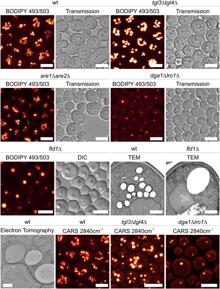
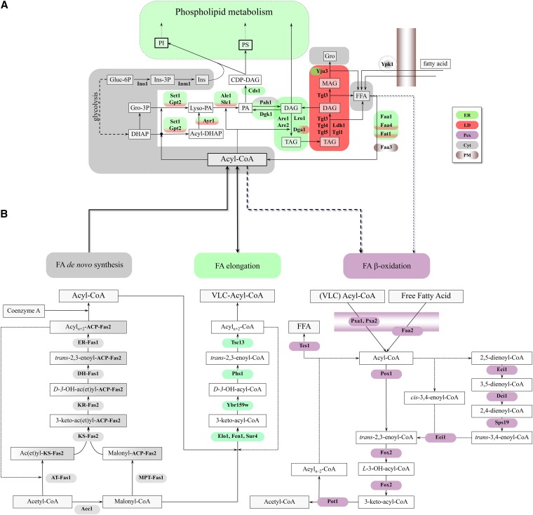
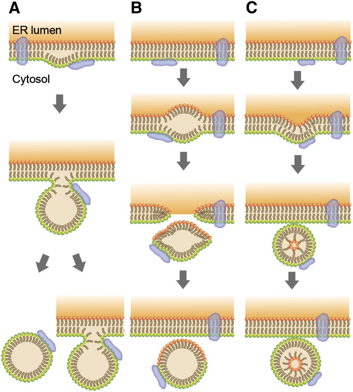
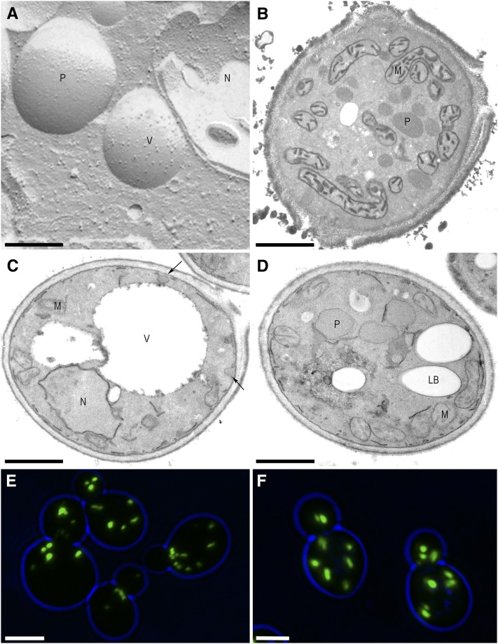
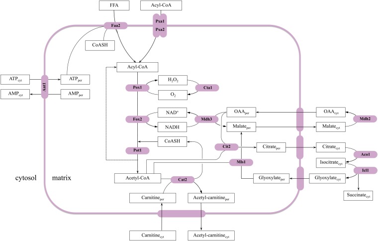
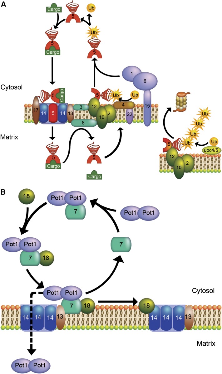
Lipid droplets (LDs) and peroxisomes are central players in cellular lipid homeostasis: some of their main functions are to control the metabolic flux and availability of fatty acids (LDs and peroxisomes) as well as of sterols (LDs). Both fatty acids and sterols serve multiple functions in the cell-as membrane stabilizers affecting membrane fluidity, as crucial structural elements of membrane-forming phospholipids and sphingolipids, as protein modifiers and signaling molecules, and last but not least, as a rich carbon and energy source. In addition, peroxisomes harbor enzymes of the malic acid shunt, which is indispensable to regenerate oxaloacetate for gluconeogenesis, thus allowing yeast cells to generate sugars from fatty acids or nonfermentable carbon sources. Therefore, failure of LD and peroxisome biogenesis and function are likely to lead to deregulated lipid fluxes and disrupted energy homeostasis with detrimental consequences for the cell. These pathological consequences of LD and peroxisome failure have indeed sparked great biomedical interest in understanding the biogenesis of these organelles, their functional roles in lipid homeostasis, interaction with cellular metabolism and other organelles, as well as their regulation, turnover, and inheritance. These questions are particularly burning in view of the pandemic development of lipid-associated disorders worldwide.


Figures

Figure 1
Figure 1
Morphological characteristics of yeast lipid droplets. (Rows 1–3, left panels) Fluorescence images of LDs that are labeled with BODIPY 493/503 (Wolinski and Kohlwein 2008; Wolinski et al. 2009a, 2012). (Right panels) Corresponding transmission images. All strains, except the fld1Δ mutant, were cultivated for 72 h in YPD complete medium; fld1Δ mutants were grown in synthetic complete (minimal) medium with 2mg/liter inositol for 12 h. Images were obtained by confocal laser scanning microscopy and represent projections of 8–12 optical sections. wt, wild type; tgl3Δ tgl4Δ, mutant lacking the major TAG lipases; are1Δ are2Δ, mutant lacking the steryl ester synthases and thus harboring LDs that contain TAG only; dga1Δ lro1Δ, mutant lacking acyl-CoA and phospholipid-dependent diacylglycerol (DAG) acyltransferases and thus harboring LDs that contain SE only; fld1Δ mutant, lacking the yeast ortholog of seipin. TEM: transmission electron microscopy images of wild type (wt) and the fld1Δ mutant. (Row 4) Electron tomography (ET) of LDs in wild type, showing close association of LDs with the ER membrane. CARS: coherent anti-Stokes Raman scattering microscopy of LDs in wild type, tgl3Δ tgl4Δ mutant, and dga1Δ lro1Δ mutant. CARS is a label-free imaging technique that generates contrast by imaging molecular vibrations at 2840 cm−1. Scale bar: 500 nm in the TEM images, 200 nm in the ET image, and 5 μm in the fluorescence/transmission images. See text for details. Images courtesy of H. Wolinski (fluorescence and CARS microscopy) and D. Kolb (electron microscopy and tomography).
Figure 2
Figure 2
(A) Metabolic pathways of TAG synthesis and degradation and their subcellular localization (adapted from Kohlwein 2010b and Henry et al. 2012). Phospholipids and TAG share DAG and PA as common precursors. In the de novo synthesis of phospholipids, PA serves as the immediate precursor of CDP-DAG, precursor to PI, PGP, and PS. PA is dephosphorylated to DAG, which serves as the precursor of PE and PC in the Kennedy pathway. DAG also serves as the precursor for TAG and can be phosphorylated, regenerating PA. The names of the enzymes that are discussed in detail in the text are shown adjacent to the arrows of the metabolic conversions in which they are involved, and the gene–enzyme relationships are listed in Table 1. Lipids and intermediates are boxed, with the most abundant lipid classes boxed by bold lines. Enzyme names are indicated in boldface type. TAG, triacylglycerols; PI, phosphatidylinositol; PA, phosphatidic acid; CDP-DAG, CDP-diacylglycerol; DAG, diacylglycerol; MAG, monoacylglycerol; Gro, glycerol; Gluc-6P, glucose-6 phosphate; DHAP, dihydroxyacetone phosphate, PS, phosphatidylserine; FFA, free fatty acids; Ins, inositol. Nucl, nucleus; ER, endoplasmic reticulum; Mito, mitochondria; LD, lipid droplets; G/E/V, Golgi, endosomes, vacuole; Pex, peroxisomes; Cyt, cytoplasma; PM, plasma membrane. See text for details. (B) Metabolic pathways of fatty acid metabolism. FA de novo synthesis and elongation: FA (type I) de novo synthesis requires the synthesis of malonyl-CoA by the acetyl-CoA carboxylase Acc1. This cytosolic trifunctional enzyme harbors a covalently bound biotin, an N-terminal biotin carboxylase domain, and a C-terminal transcarboxylase domain (Tehlivets et al. 2007). Malonyl-CoA is used by the cytosolic FA synthase complex, consisting of Fas1 (β-subunit) and Fas2 (α-subunit), which are organized in a hexameric α6β6 complex. Fas1 harbors acetyl transferase (AT), enoyl reductase (ER), dehydratase (DH), and malonyl-palmitoyl transferase (MPT) activities; Fas2 contains the acyl carrier protein (ACP), 3-ketoreductase (KR), 3-ketosynthase (KS), and phosphopantheine transferase activities. The product of FA synthesis in yeast is acyl-CoA, typically C14–C16 carbon atoms in length (Tehlivets et al. 2007). Activated FAs may be elongated to VLCFAs by the activity of Elo1, Fen1/Elo2, and Sur4/Elo3 (condensing enzymes); Ybr159w (reductase); Phs1 (dehydratase); and Tsc13 (enoyl-CoA reductase). Yeast also expresses a set of bacterial type II enzymes (as individual polypeptides) that perform the same reactions in mitochondria, but are encoded by nuclear genes (Tehlivets et al. 2007). Mitochondrial FA synthesis presumably generates FA only up to C8, which is a precursor for lipoic acid synthesis. FAs are degraded by β-oxidation. β-oxidation in yeast occurs exclusively in peroxisomes. Medium chain fatty acids enter peroxisomes as free fatty acids (FFA) and are activated by a peroxisomal acyl-CoA synthetase, Faa2. ATP that is required for this activation step is imported into the organelle via Ant1. Long chain fatty acids, such as oleate, are activated outside the organelle by Fat1, Faa1, or Faa4 and taken up as CoA esters (acyl-CoA) via a peroxisomal ABC transporter that consists of the heterodimer Pxa1/Pxa2. Inside peroxisomes, CoA esters undergo dehydrogenation by Pox1, hydratation/dehydrogenation by Fox2, and ultimately thiolytical cleavage by Pot1, leading to acetyl-CoA and an acyl-chain shortened by two carbon atoms. Hydrogen peroxide produced by Pox1 is degraded by peroxisomal catalase T, Cta1. NADH is exported to the cytosol via a malate shuttle that involves peroxisomal (Mdh3) and cytosolic (Mdh2) malate dehydrogenases. The transporter for malate and oxaloacetate has not been identified yet. Acetyl-CoA is transported to the cytosol via carnitine-dependent acetyl-CoA transport (involving Cat2) or via the glyoxylate cycle (see Figure 5). Unsaturated FAs, such as oleic acid with the double bond between C9 and C10, can be fully oxidized only in the presence of auxiliary enzymes, but the precise mechanism is controversial. Eci1 is a Δ3,Δ2-enoyl-CoA isomerase in the so-called isomerase-dependent major pathway, which catalyzes the positional and stereochemical isomerization of cis-3-enoyl-CoA to trans-2-enoyl-CoA; this reaction is required after oleic acid (as coenzyme A derivative) has been shortened by three rounds of β-oxidation, since only trans-2-enoyl-CoA is a β-oxidation substrate. Eci1 also isomerizes a fraction of 2-trans, 5-cis-dienoyl-CoA to 3,5-dienoyl-CoA, which has two conjugated double bonds in trans (3) and cis (5) configuration. This compound is presumably degraded by the minor pathway that involves Dci1, Sps19, and Eci1. Alternatively, 3,5-dienoyl-CoA is hydrolyzed by Tes1 thioesterase-dependent pathway) to the free FA and coenzyme A.
Figure 3
Figure 3
Models of lipid droplet biogenesis (adapted from Guo et al. (2009). (A) According to the “lensing model,” neutral lipids are deposited between the leaflets of the ER membrane: after reaching a critical size, the neutral lipid core bulges out and the LD is formed; the LD surface monolayer is derived solely from the cytosolic leaflet of the ER membrane. Subsequently, the LD may completely separate from the ER membrane, or remain attached, with the surface layer forming a continuum with the ER. (B) Bicelle formation: LD formation similar to model in A, but the LD is excised from the ER membrane, and both ER membrane leaflets contribute to the LD surface monolayer. (C) Vesicle formation. Inclusion of the neutral lipid core in the membrane vesicle requires rearrangement of the inner leaflet of the bilayer. These models explain the origin of the phospholipid membrane, which stems either from the cytoplasmic leaflet or from both leaflets of the ER membrane, respectively. Unclear is what limits the expansion of the neutral lipid core between the leaflets, what determines the orientation of LD extrusion toward the cytosol, and how the integrity of the ER membrane is maintained. Notably, none of the intermediate stages representing neutral lipid deposits between the ER membrane leaflets, nascent lipid droplets in the ER, or lipid-filled vesicular structures have been experimentally observed in wild-type cells.
Figure 4
Figure 4
Morphological characteristics of yeast peroxisomes. (A) Freeze-etch replica of oleic acid-grown S. cerevisiae cells showing the fraction faces of the different organelles. Peroxisomes contain very smooth fracture faces, indicative of a low abundance of integral membrane proteins. (B) Thin section of a cell grown on oleic acid cytochemically stained for catalase activity, using diamino-benzidine and hydrogen peroxide. In these cells, numerous stained, electron-dense peroxisomes are present. Staining of the mitochondrial cristae is due to cytochrome c peroxidase activity. Ultrathin sections of (C) glucose-grown and (D) oleic acid-grown and KMnO4-fixed S. cerevisiae G910 cells (Veenhuis et al. 1987). The cell grown on glucose displays only a few very small peroxisomes (arrows), whereas strong peroxisome proliferation is evident in the oleic acid-grown cell. (E and F) Fluorescence microscopy of (E) glucose-grown and (F) oleic acid-grown cells expressing GFP-SKL to label peroxisomes in wild-type strain BY4742. Notably, in this strain background, the difference in peroxisome number on glucose and oleic acid media is far less pronounced compared to strain G910. N, nucleus; M, mitochondrion; P, peroxisome; V, vacuole; LD, lipid droplet. Scale bar: 200 nm in A, 1 μm in B–D, and 3 μm in E and F.
Figure 5
Figure 5
Compartmentalization of the β-oxidation pathway and the glyoxylate cycle. The enzymes of the β-oxidation (see Figure 2B) as well as key enzymes of the glyoxylate cycle are localized to peroxisomes in S. cerevisiae. In addition to the β-oxidation enzymes, three peroxisomal membrane-associated proteins are required for fatty acid oxidation in peroxisomes, namely the ABC transporter Pxa1/Pxa2 for the import of long-chain acyl-CoA, Faa2 for the activation of medium chain FAs, and the transporter Ant1 for import of ATP. The glyoxylate cycle converts two acetyl-CoA molecules into succinate and contributes to the export of acetyl-CoA that is produced in the β-oxidation cycle. Glyoxylate and the first acetyl-CoA molecule are condensed by malate synthase (Mls) to malate; malate dehydrogenase (Mdh) converts malate to oxaloacetate (OAA); and isocitrate synthase (Cit) condenses OAA and a second acetyl-CoA molecule to form citrate. Aconitase (Aco) catalyzes the isomerization of citrate into isocitrate, which is cleaved by isocitrate lyase (Icl) into succinate and glyoxylate. Glyoxylate can be used for the next round of the glyoxylate cycle, whereas succinate is used to replenish the citric acid cycle or to function as a precursor for amino acid or carbohydrate biosynthesis. In S. cerevisiae, citrate synthase, Cit2, and malate synthase Mls1 are peroxisomal enzymes, whereas Icl1 is cytosolic. This is in contrast to plants, filamentous fungi, and other yeast species, in which Icl is peroxisomal as well. Acetyl-CoA can also be exported via the carnitine shuttle, which involves peroxisomal Cat2. The malate shuttle is responsible for NADH export. The predicted small molecule transporters involved in both shuttles or in the glyoxylate cycle have not been identified yet.
Figure 6
Figure 6
Peroxisomal protein import. (A) Hypothetical model of PTS1 protein import. First, cytosolic Pex5 binds a newly synthesized PTS1-containing cargo protein (“cargo”). The PTS1 binds to the C-terminal TPR domain of Pex5. Next, Pex5 docks to the receptor-docking complex at the peroxisomal membrane, which is composed of Pex13, Pex14, and Pex17. Docking involves the N-terminal domain of Pex5, indicated as a spiral. Subsequently, the Pex5-cargo complex is imported into the organellar matrix. Pex5 most likely forms a transient pore in the peroxisomal membrane. The Pex5-cargo complex than dissociates in a process that involves Pex8, a peripheral membrane protein in the peroxisomal matrix. Finally, Pex5 is recycled back to the cytosol, a process that enables it to bind the next PTS1 cargo protein. Recycling involves mono-ubiquitination of Pex5 by the UBC protein Pex4, which is recruited to the peroxisomal membrane by Pex22. The three RING finger proteins Pex2, Pex10, and Pex12 are proposed to serve as E3 ligases. The ubiquitinated Pex5 is pulled out of the membrane by the AAA proteins Pex1 and Pex6, which are associated with the peroxisomal membrane via Pex15. When Pex5 recycling fails, it becomes polyubiquitinated by Ubc4/5 and degraded by the proteasome. (B) Hypothetical model of PTS2 protein import. Dimeric Pot1 is shown as an example of a typical PTS2 protein. Dimeric Pot1 first binds to the PTS2 receptor Pex7. Subsequently, the coreceptor Pex18 (and possibly also Pex21) binds to the receptor/cargo complex. Pex7 associates with Pex13 of the docking complex. After import of the Pot1 cargo into peroxisomes by an as-yet-unknown mechanism Pex7 recycles back to the cytosol. Pex18, however, first forms a complex with Pex14. Whether and how Pex18 recycles for another round of import is unknown.

References

    1. AbdelRaheim S. R., Cartwright J. L., Gasmi L., McLennan A. G., 2001. The NADH diphosphatase encoded by the Saccharomyces cerevisiae NPY1 nudix hydrolase gene is located in peroxisomes. Arch. Biochem. Biophys. 388: 18–24 - PubMed
    1. Adeyo O., Horn P. J., Lee S., Binns D. D., Chandrahas A., et al. , 2011. The yeast lipin orthologue Pah1p is important for biogenesis of lipid droplets. J. Cell Biol. 192: 1043–1055 - PMC - PubMed
    1. Agarwal A. K., Garg A., 2003. Congenital generalized lipodystrophy: significance of triglyceride biosynthetic pathways. Trends Endocrinol. Metab. 14: 214–221 - PubMed
    1. Agarwal A. K., Barnes R. I., Garg A., 2004. Genetic basis of congenital generalized lipodystrophy. Int. J. Obes. Relat. Metab. Disord. 28: 336–339 - PubMed
    1. Agne B., Meindl N. M., Niederhoff K., Einwachter H., Rehling P., et al. , 2003. Pex8p: an intraperoxisomal organizer of the peroxisomal import machinery. Mol. Cell 11: 635–646 - PubMed

Publication types