. 2013 Feb 1;41(4):2416-27.
doi: 10.1093/nar/gks1345. Epub 2012 Dec 28.

DNA target sequence identification mechanism for dimer-active protein complexes

Affiliations

DNA target sequence identification mechanism for dimer-active protein complexes

Markita P Landry et al. Nucleic Acids Res. .

Abstract

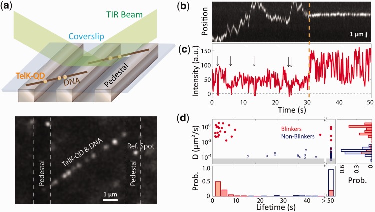
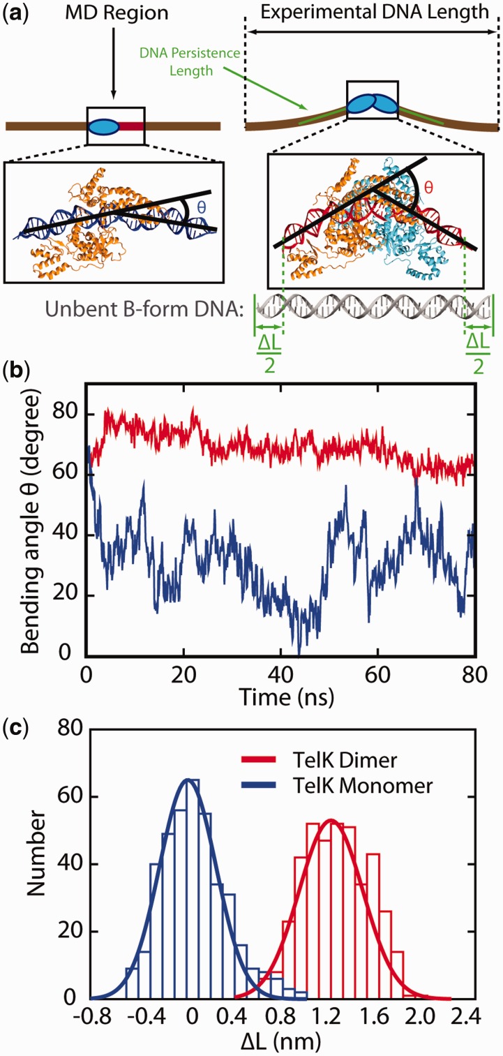
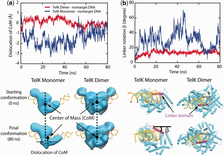
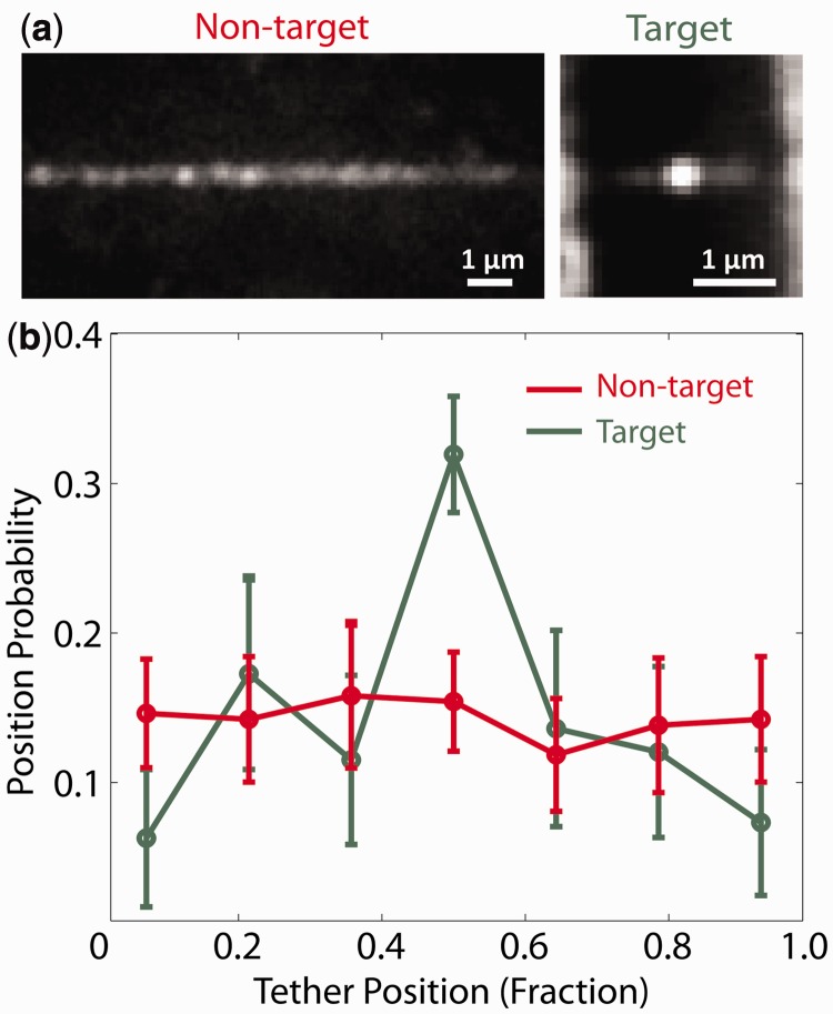
Sequence-specific DNA-binding proteins must quickly and reliably localize specific target sites on DNA. This search process has been well characterized for monomeric proteins, but it remains poorly understood for systems that require assembly into dimers or oligomers at the target site. We present a single-molecule study of the target-search mechanism of protelomerase TelK, a recombinase-like protein that is only active as a dimer. We show that TelK undergoes 1D diffusion on non-target DNA as a monomer, and it immobilizes upon dimerization even in the absence of a DNA target site. We further show that dimeric TelK condenses non-target DNA, forming a tightly bound nucleoprotein complex. Together with theoretical calculations and molecular dynamics simulations, we present a novel target-search model for TelK, which may be generalizable to other dimer and oligomer-active proteins.


Figures

Figure 1.
Figure 1.
TelK monomers diffuse along non-target DNA, whereas dimers immobilize. (a) Schematic of TIRFM experimental set-up and representative fluorescence image. An etched glass slide with 1 × 1 µm pedestals separated by 7-µm etches was coated with neutravidin. Dual-biotinylated λ-DNA was flowed in to form DNA bridges, and the chamber was, subsequently, incubated with QD-labelled TelK monomers (here, 340 nM). QDs stuck to the glass surface were used as reference spots to ensure that drift and background motion were minimal. TelK concentrations used in all TIRF experiments ranged from 70 to 1350 nM. (b) Kymograph of QD-labelled TelK on λ-DNA showing mobile (1–31 s) and stationary (32–50 s) states after analysis and background subtraction with Gaussian fitting of spots. (c) Fluorescence intensity corresponding to the kymograph. The intensity of the mobile TelK doubles as it becomes immobile along the λ-DNA bridge. QD-blinking events (arrows), known to occur for single QDs, are observed only before TelK immobilization. (d) Diffusion coefficient and lifetime for blinking fluorescence spots (n = 114, red) and non-blinking spots (n = 81, blue). The shaded area represents the limit of sensitivity of our assay. The distribution of diffusion coefficients (right panel) is bimodal, with blinkers diffusing approximately four orders of magnitude faster than non-blinkers. As shown in the lifetime distributions (bottom panel), blinking spots also remained DNA-bound for shorter times (4.2 s) than non-blinkers (>50 s).
Figure 2.
Figure 2.
TelK dimers condense non-target DNA. (a) Schematic of optical trap experimental set-up. A DNA tether was formed in a stream containing buffer only and, subsequently, moved into a stream with buffer containing TelK. TelK binding to the DNA tether causes DNA condensation. (b) Time traces showing single DNA condensation events for tethers exposed to 100 nM TelK (blue) fit with a step-finder algorithm (red), and protein-free control trace (grey). Three characteristic times are highlighted in a sample trace: the time to first condensation, the condensed state lifetime and uncondensed state time. Traces offset for clarity. (c) The time for first TelK-induced condensation (green) is strongly dependent on (TelK), whereas the dwell time of the condensed state (blue) and the time between condensation events (red) do not depend on (TelK). (d) Normalized histogram of step size (Δx) for all observed condensation events (n = 93) because of DNA-induced TelK bending, with Gaussian fit to the condensation step size expected from the TelK dimer-DNA crystal structure.
Figure 3.
Figure 3.
MD simulations confirm that Telk dimers bend non-target DNA, whereas monomers cannot. (a) Schematic representation of DNA condensation as would be measured in optical trap experiments (brown line) and MD simulations (boxed region). ΔL is defined as the difference between the contour length and the end-to-end distance of the 44-bp DNA substrate. MD snapshots of a TelK dimer-DNA complex and a TelK monomer-DNA complex are shown. DNA adopts a bent conformation in interaction with the TelK dimer, but not with the TelK monomer. (b) DNA bend angle induced by a TelK dimer (red) and a TelK monomer (blue). (c) Distribution of the change in end-to-end distance in the 44-bp MD DNA substrate (ΔL) induced by a TelK dimer and monomer in MD simulations.
Figure 4.
Figure 4.
TelK monomers (blue) are more mobile on non-target DNA compared to dimers (red). (a) Dislocation of TelK dimer and monomer along non-target DNA axis based on MD simulations. (b) Rotation of TelK linker domain (SER208-ASN229). The snapshots show the starting and final positions of the linker domain (magenta) in TelK monomer and dimer after 80 ns. DNA is shown in yellow, and TelK is shown in cyan.
Figure 5.
Figure 5.
TelK binds preferentially to target DNA. (a) Average TelK fluorescent spot positions along target (green, n = 182) and non-target (red, n = 253) DNA, obtained by aligning and overlapping fluorescence images. TelK concentration range used in all TIRF experiments ranged from 70 to 1350 nM. (b) Position probability distribution of TelK along target (green) and non-target (red) DNA.
Figure 6.
Figure 6.
Kinetics of TelK-induced dimerization and target search. (a) Model of TelK target search. TelK monomers in solution bind DNA with rate kon and scan rapidly along non-target DNA with mean diffusion coefficient Dmonomer = 1.8 µm2/s. Monomers localize the target site with rate ktarget and bind tightly or dissociate from non-target DNA with average rate koff = 0.24 s−1. Preferential and stable binding of a TelK monomer allows a second monomer to dimerize at the target site and form a kinked DNA–TelK complex primed for catalysis. Occasionally, mobile monomers encounter each other on non-target DNA with rate kdimer and form stable, immobile dimers (Ddimer < 1 × 10⁻⁴ µm2/s) that ‘test’ their substrate by condensing the DNA transiently. Eventually, dimers on non-target DNA dissociate or separate into mobile monomers again (rate < 0.01 s−1). (b) Implementation of kinetic model in (a) via stochastic simulations using experimentally derived kinetic parameters. Average rates for first dimerization kdimer (blue, black), and first target localization ktarget (red) as a function of average protein occupancy, <N>, on DNA. For mean occupancies >1, target-finding and dimerization rates obey simple scaling laws of N2 and N4, respectively. For mean occupancies <1, dimerization follows of N2 scaling. Provided TelK occupancy is reasonably small, target localization consistently occurs faster than dimerization. Simulated rates are in good agreement with experimentally derived rates of first dimerization.

Similar articles

Cited by

References

    1. Koslover EF, Diaz de la Rosa MA, Spakowitz AJ. Theoretical and computational modeling of target-site search kinetics in vitro and in vivo. Biophys. J. 2011;101:856–865. - PMC - PubMed
    1. van den Broek B, Lomholt MA, Kalisch SMJ, Metzler R, Wuite GJL. How DNA coiling enhances target localization by proteins. Proc. Natl Acad. Sci. USA. 2008;105:15738–15742. - PMC - PubMed
    1. Gowers DM, Wilson GG, Halford SE. Measurement of the contributions of 1D and 3D pathways to the translocation of a protein along DNA. Proc. Natl Acad. Sci. USA. 2005;102:15883–15888. - PMC - PubMed
    1. Ramanathan SP, van Aelst K, Sears A, Peakman LJ, Diffin FM, Szczelkun MD, Seidel R. Type III restriction enzymes communicate in 1D without looping between their target sites. Proc. Natl Acad. Sci. USA. 2009;106:1748–1753. - PMC - PubMed
    1. Halford SE, Marko JF. How do site-specific DNA-binding proteins find their targets? Nucleic Acids Res. 2004;32:3040–3052. - PMC - PubMed

Publication types