. 2013 Feb 1;41(4):2673-82.
doi: 10.1093/nar/gks1303. Epub 2012 Dec 28.

Crystal structure of an intermediate of rotating dimers within the synaptic tetramer of the G-segment invertase

Affiliations

Crystal structure of an intermediate of rotating dimers within the synaptic tetramer of the G-segment invertase

Christopher J Ritacco et al. Nucleic Acids Res. .

Abstract

The serine family of site-specific DNA recombination enzymes accomplishes strand cleavage, exchange and religation using a synaptic protein tetramer. A double-strand break intermediate in which each protein subunit is covalently linked to the target DNA substrate ensures that the recombination event will not damage the DNA. The previous structure of a tetrameric synaptic complex of γδ resolvase linked to two cleaved DNA strands had suggested a rotational mechanism of recombination in which one dimer rotates 180° about the flat exchange interface for strand exchange. Here, we report the crystal structure of a synaptic tetramer of an unliganded activated mutant (M114V) of the G-segment invertase (Gin) in which one dimer half is rotated by 26° or 154° relative to the other dimer when compared with the dimers in the synaptic complex of γδ resolvase. Modeling shows that this rotational orientation of Gin is not compatible with its being able to bind uncleaved DNA, implying that this structure represents an intermediate in the process of strand exchange. Thus, our structure provides direct evidence for the proposed rotational mechanism of site-specific recombination.


Figures

Figure 1.
Figure 1.
The mechanism of Gin inversion. Top: bar scheme depicting the arrangement of the phage Mu DNA prior to and after a successful inversion event. Bottom: a dimer of Gin binds to each gix site (data not shown) and forms a homo-dimeric tetramer at synapsis. The activating factor Fis (data not shown) causes a conformational change in Gin allowing catalysis. The cleavage reactions result in a double-strand break covalently linked to the protein through a phosphodiester bond at the gix site. The strands are exchanged through 180° of rotation of the DNA about the exchange interface and then are religated to complete the inversion.
Figure 2.
Figure 2.
The structure of the synaptic tetramer of Gin. (A) The final 2Fo–Fc electron density map, calculated at 4.0 Å and contoured at 1σ, is superimposed onto the subunit structure of Gin within one asymmetric unit. (B and C) Two orthogonal views of the tetrameric synaptic complex of Gin are displayed to illustrate the difference in interfaces in the synaptic tetramer. The subunit dimer-AB and the subunit dimer-CD create the flat exchange interface of Gin. (D) The exchange interface (the x and y plane) is exclusively created by the E helices. The synaptic interface between subunits A/B or subunits C/D is created by both the E and D helices. The three orthogonal 222-axes (arbitrarily designated here as x,y,z), N and C termini, some helices and subunits A (yellow), B (magenta), C (blue) and D (green) are labeled.
Figure 3.
Figure 3.
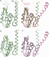
The conformation of the subunit of Gin. (A) Superposition of the NTD of Gin (green) onto the NTD of a subunit of the synaptic structure of γδ resolvase (cyan). (B) Superposition of the E helices of Gin and the synaptic γδ resolvase. (C) Superposition of the NTD of Gin onto a subunit of the Site I dimer of γδ resolvase (magenta). (D) Superposition of the NTDs of a subunit from the dimer and tetramer structures of γδ resolvase.
Figure 4.
Figure 4.
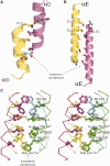
The synaptic and exchange interfaces of the synaptic complex of Gin. (A and B) The putative contacts made between the D (A) and E (B) helices at the synaptic interface. At the interface between the D helices, a simple modeling of an alternate rotamer of E79 (cyan A) creates a salt bridge between E79 and K71. (B) The modeled residues at the synaptic interface between the E helices may interact through electrostatic, hydrogen bonds and salt bridges between E117 and R102. (C) Stereo view of the exchange interface of Gin highlighting the hydrophobic residues at its core.
Figure 5.
Figure 5.
The subunit rotation model at the exchange interface of the serine recombinases. (A) Cartoon illustration of the rotation of subunits around the exchange interface. (B) Three synaptic tetramers with emphasis on the orientation of their E Helices [Sin (magenta), Resolvase (cyan) and Gin (green)]. The appropriate position of each tetramer on the pathway of subunit rotation is shown. (C) The cross angles of the E Helices of the three synaptic tetramers shown perpendicular to (B). The arrows indicate the direction (N to C) of the sequence in each E Helix.

References

    1. Craig NL. Mobile DNA II. Washington, D.C: ASM Press; 2002.
    1. Guo F, Gopaul D, van Duyne G. Structure of Cre recombinase complexed with DNA in a site-specific recombination synapse. Nature. 1997;389:40–46. - PubMed
    1. Gibb B, Gupta K, Ghosh K, Sharp R, Chen J, Van Duyne GD. Requirements for catalysis in the Cre recombinase active site. Nucleic Acids Res. 2010;38:5817–5832. - PMC - PubMed
    1. Ghosh K, Lau C, Gupta K, Van Duyne G. Preferential synapsis of loxP sites drives ordered strand exchange in Cre-loxP site-specific recombination. Nat. Chem. Biol. 2005;1:275–282. - PubMed
    1. Guo F, Gopaul D, Van Duyne G. Asymmetric DNA bending in the Cre-loxP site-specific recombination synapse. Proc. Natl Acad. Sci. USA. 1999;96:7143–7148. - PMC - PubMed

Publication types