. 2013 Feb 1;41(4):2594-608.
doi: 10.1093/nar/gks1361. Epub 2012 Dec 28.

RNA dimerization plays a role in ribosomal frameshifting of the SARS coronavirus

Affiliations

RNA dimerization plays a role in ribosomal frameshifting of the SARS coronavirus

Daniella Ishimaru et al. Nucleic Acids Res. .

Abstract

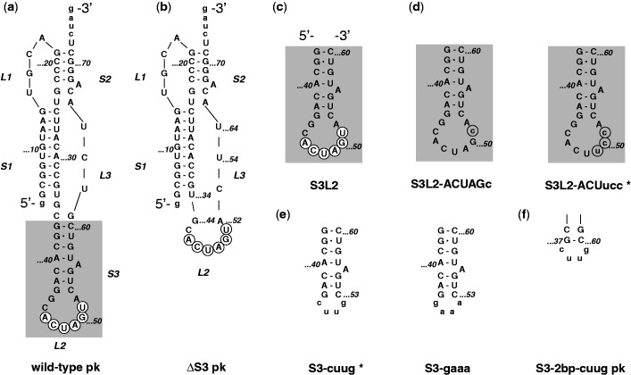
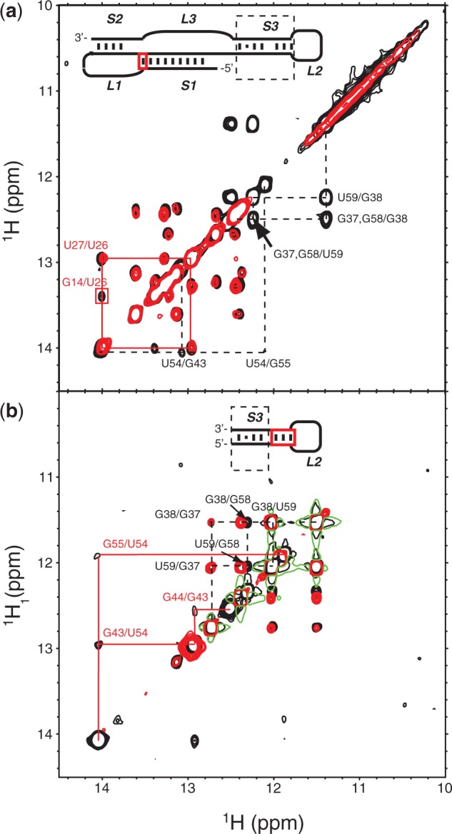
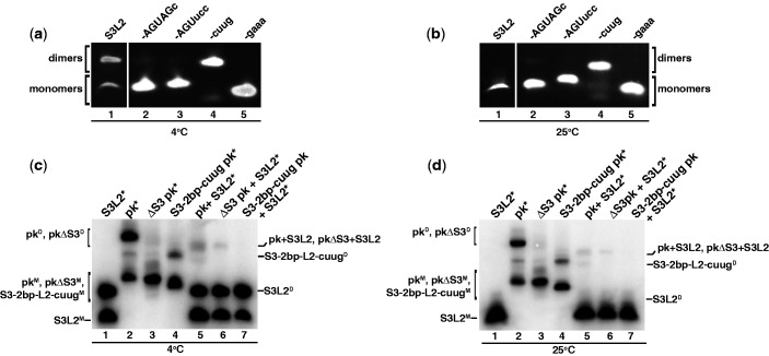
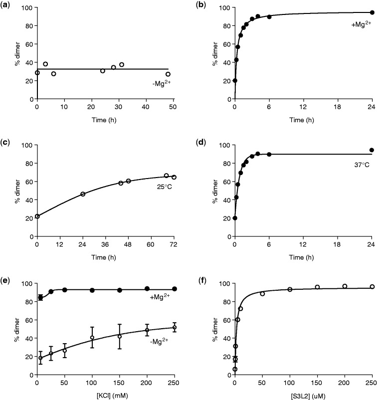
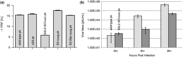
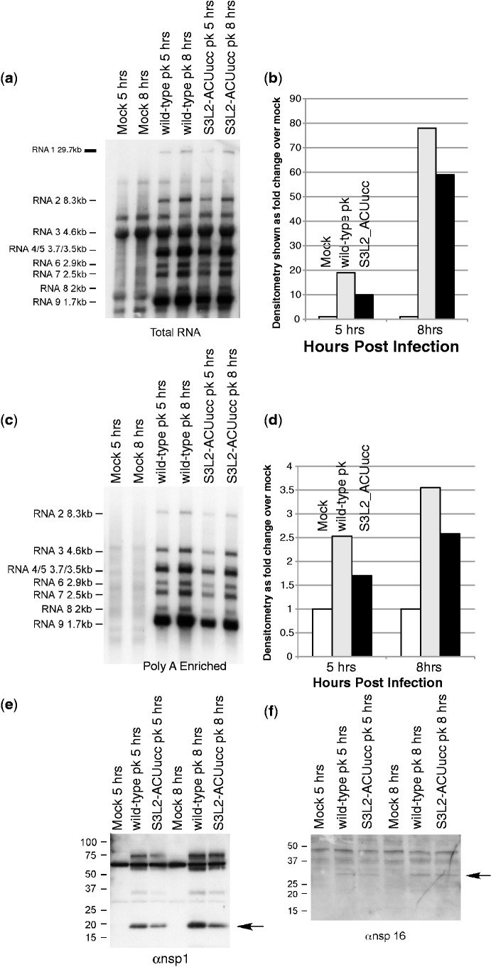
Messenger RNA encoded signals that are involved in programmed -1 ribosomal frameshifting (-1 PRF) are typically two-stemmed hairpin (H)-type pseudoknots (pks). We previously described an unusual three-stemmed pseudoknot from the severe acute respiratory syndrome (SARS) coronavirus (CoV) that stimulated -1 PRF. The conserved existence of a third stem-loop suggested an important hitherto unknown function. Here we present new information describing structure and function of the third stem of the SARS pseudoknot. We uncovered RNA dimerization through a palindromic sequence embedded in the SARS-CoV Stem 3. Further in vitro analysis revealed that SARS-CoV RNA dimers assemble through 'kissing' loop-loop interactions. We also show that loop-loop kissing complex formation becomes more efficient at physiological temperature and in the presence of magnesium. When the palindromic sequence was mutated, in vitro RNA dimerization was abolished, and frameshifting was reduced from 15 to 5.7%. Furthermore, the inability to dimerize caused by the silent codon change in Stem 3 of SARS-CoV changed the viral growth kinetics and affected the levels of genomic and subgenomic RNA in infected cells. These results suggest that the homodimeric RNA complex formed by the SARS pseudoknot occurs in the cellular environment and that loop-loop kissing interactions involving Stem 3 modulate -1 PRF and play a role in subgenomic and full-length RNA synthesis.


Figures

Figure 1.
Figure 1.
SARS constructs. (a) The three-stemmed wild-type SARS pseudoknot. Stems are labeled S1, S2 and S3 in the order that they occur 5′ to 3′ along the RNA. Accordingly, loops are labeled L1, L2 and L3. Note that L1 and L3 join adjacent stems, while L2 closes S3 (highlighted using gray box). Only the last two digits of the wild-type sequence numbering are used for clarity. The palindromic sequence 5′-ACUAGU-3′ embedded into L2 is indicated using white circles. Dashes represent Watson–Crick and the dot G•U Wobble base-pairing as confirmed by NMR spectroscopy. (b) Stem 3 deletion mutant ΔS3 pk. (c) S3L2 hairpin construct S3L2 spanning nucleotides G37 to C60. (d) S3L2 hairpin constructs S3L2-ACUucc and S3L2-ACUAGc with L2 mutations that render the palindromic sequence asymmetrical (highlighted using gray circles) while conserving a Serine codon. (e) S3L2 hairpin constructs S3-cuug and S3-gaaa where the 9 nt L2 is replaced with the smaller tetraloops 5′-cuug-3′ and 5′-gaaa-3′, respectively. (f) SARS pseudoknot variants S3-2 bp-cuug pk with a shortened Stem 3 is capped with a 5′-cuug-3′ tetraloop. S3-cuug and S3L2-ACUucc variant constructs highlighted with an asterisk (*) were also generated in the context of full-length pk.
Figure 2.
Figure 2.
NMR secondary structure comparison of wild-type SARS-CoV pk, ΔS3 pk, S3 and S3L2-ACUucc mutants. (a) Imino regions of 2D 1H,1H-NOESY experiments collected on wild-type SARS-CoV pk (black contours) and ΔS3 pk mutant (red contours), respectively. Dashed black lines show the imino proton walk in the S3 stem. The base-paired region of S3 is deleted in the ΔS3 pk mutant; however, L2 is left intact. Solid red lines show the sequential NOE correlations involving the imino proton U26 located in S1, and the red box highlights the cross peak connecting imino protons U26 and G14 adjacent to the S1-S2 junction, which is absent in the ΔS3 pk mutant. Only the last two digits of the wild-type sequence numbering are used for clarity. The schematic SARS-CoV pk inset highlights the corresponding S3 stem (dashed box) as well as the G14-C25 basepair location in S1 (solid red box). (b) Imino regions of 2D 1H,1H-NOESY experiments collected on wild-type SARS S3 (black contours) and the S3L2-ACUucc mutant (red contours), respectively. Dashed black lines show the imino proton walk in the lower portion of the S3 stem. Solid red lines highlight the sequential cross peaks in the upper portion of S3 correlating imino protons G55, U54 and G43 adjacent to L2, which are broadened beyond detection in the S3L2-ACUucc mutant. The schematic SARS S3L2 inset highlights the corresponding lower S3 stem (dashed box) as well as the base-paired region in the upper S3 stem (solid red box).
Figure 3.
Figure 3.
Detection of SARS RNA dimers. RNA transcripts (150 µM/each) were incubated at 37°C for 30 min in the presence of 200 mM KCl without MgCl2. Samples were separated on a 10% native PAGE. (a) Native gel analysis at 4°C of S3L2-only variants: S3L2, S3L2-ACUAGc, S3L2-ACUucc, S3-cuug and S3-gaaa. Gels were stained with ethidium bromide and visualized by UV. S3L2 transcript was independently analysed. (b) Same as in (a) but at 25°C. (c) Native gel analysis at 4°C of the following transcripts: wild-type S3L2, wild-type pk, Stem 3 deletion mutant ΔS3 pk and S3-2 bp-cuug. RNA variants were incubated with 32P-labeled transcripts in trace [1 pM] indicated by *. (d) Same as in (c), but at 25°C. For (c) and (d) samples were separated on a 10% native PAGE, which was dried and analysed by phosphorimaging. Uppercase ‘M’ denotes monomer and uppercase ‘D’, dimer.
Figure 4.
Figure 4.
Evaluation of dimer-promoting conditions for SARS S3L2. Wild-type SARS S3L2 transcripts (150 µM) were incubated in 10 mM Na2HPO4, pH 6.5, 0.5 mM EDTA, 100 mM KCl and 5 mM MgCl2 for 6 h at 37°C, unless otherwise stated. Samples were separated on a native PAGE in Tris Borate buffer. Gels were dried and analysed by phosphorimaging. (a) RNAs were incubated in the absence of MgCl2. Aliquots were removed at the following time-points: 0 h, 3 h, 6 h, 24 h, 28 h, 31 h and 48 h. (b) Same as in (a) but in the presence of 5 mM MgCl2. Aliquots were removed at the following time-points: 0 min, 15 min, 30 min, 1 h, 1:30 h, 2 h, 3 h, 4 h, 6 h and 24 h. (c) RNAs were incubated at 25°C in the presence of 5 mM MgCl2. Aliquots were removed at the following time-points: 0 h, 24 h, 44 h, 48 h, 68 h and 72 h. (d) Same as in (c) but at 37°C. Aliquots were removed at the following time-points: 0′, 15′, 30′, 1 h, 1:30 h, 2 h, 3 h, 4 h, 6 h and 24 h. (e) RNAs were incubated in the absence (open symbols) or in the presence of 5 mM MgCl2 (closed symbols) and varying concentrations of KCl (6–250 mM). (f) RNAs (0.1–250 µM) were incubated at 37°C for 6 h in the presence of MgCl2.
Figure 5.
Figure 5.
Schematic structures of SARS RNA dimers. (a) Stem–loop sequence and secondary structure for Stem 3 from SARS-CoV (S3L2M). (b) Schematic representation of the Stem 3 loop–loop kissing homodimer, with individual stem–loops shown using black and gray letters, respectively (S3L2D*). (c) Schematic of an extended S3 duplex, with individual stem–loops shown using black and gray letters, respectively (S3L2D). (d) Stem–loop sequence and secondary structure for the 5′-cuug-3′-capped Stem 3 variant (S3-cuugM). (e) Schematic of an extended S3-cuug duplex, with individual stem–loops shown using black and gray letters, respectively (S3-cuugD).
Figure 6.
Figure 6.
Identification of the crosslinking site of the S3L2 dimer. (a) Partial alkaline hydrolysis of control S3L2 and crosslinked S3L2 [32P]ATP-RNAs. Lane 1, RNase T1 digestion of control S3L2 RNA; locations of Gs are shown on the right side. Arrow, position of C…45, the first nucleotide with reduced intensity in the partial hydrolysis. Lane 2, alkaline hydrolysis ladder. Lanes 3 and 4, partial alkaline hydrolysis of crosslinked and non-crosslinked samples, respectively. (b) Ratio of intensities from the partial hydrolysis of lanes 3 and 4 in Panel (a). Values for each lane were normalized and the intensities of each band of crosslinked over control RNAs were plotted. Error bars indicate the values obtained in four independent experiments.
Figure 7.
Figure 7.
NMR real-time monitoring of salt-dependent loop–loop kissing formation of wild-type SARS-CoV S3L2 stem–loop. (a) Time course of imino regions of 1D 1H-jump-return echo experiments. Data were collected on a SARS S3L2 sample containing 0.25 mM RNA in 500 µl volume of NMR buffer. Spectra were recorded at 295 K on a Bruker Avance III 600 (S3) MHz spectrometer. Loop–loop kissing formation was induced by spiking the NMR buffer [10 mM sodium phosphate (pH 6.5), 500 µM EDTA, 50 µM sodium azide, 9:1 H2O:D2O] with 125 mM potassium chloride. Dashed black lines follow assigned imino proton resonances at various time points. Only the last two digits of the wild-type sequence numbering are used for clarity. (b) Schematic S3L2 inset showing the loop–loop kissing geometry highlighting the lower S3 stem (black dashed box), the base-paired region in the upper S3 stem (solid red box) as well as intermolecular loop–loop kissing interactions (solid blue lines).
Figure 8.
Figure 8.
Functional analysis of wild-type SARS-CoV and S3L2-ACUucc pk mutants. (a) Frameshifting Frequencies of SARS S3L2 Pseudoknot Mutants. -1PRF (%) efficiency was determined as described in the methods. -1 PRF efficiency for ΔS3 pk is included for comparison and was reported previously (1). (b) Cultures of VeroE6 cells were inoculated with wild-type or S3L2-ACUucc pk mutant SARS-CoV at an MOI 1. Samples were taken at the indicated times and viral titers determined by plaque assay. Titers are indicated as plaque forming units/ml (ml). Error bars represent the standard deviations of three measurements. Student’s t-test: for 0 h, P = 0.6184; for 5 h, P = 0.0310; for 8 h, P = 0.0015.
Figure 9.
Figure 9.
S3L2-ACUucc SARS-CoV mutant RNA synthesis and protein expression kinetics. Cultures of VeroE6 cells were infected with wild-type or S3L2-ACUucc mutant SARS-CoV at an MOI of 1 or mock-infected. Representative northern blots with sgRNA and gRNA species and molecular weights indicated by arrows. (a) probed total RNA to visualize genomic and subgenomic viral RNA and (c) probed polyA enriched RNA to visualize the sgRNA species. Delays in RNA synthesis kinetics in S3L2-ACUucc frameshift mutant. Bars represent the densitometry measurements of fold change over mock for mock-infected, wild-type and S3L2-ACUucc SARS-CoV mutant RNA at the indicated times pi. (b) Densitometry of gRNA. (d) Densitometry of sgRNA. ORF1a versus ORF1b protein expression kinetics in S3L2-ACUucc frameshift mutant. Proteins were separated on polyacrylamide gels and probed with rabbit sera directed against (e) the replicase proteins nsp1 or (f) nsp16 as indicated. Size markers are indicated to the left of each blot.

References

    1. Plant EP, Perez-Alvarado GC, Jacobs JL, Mukhopadhyay B, Hennig M, Dinman JD. A three-stemmed mRNA pseudoknot in the SARS coronavirus frameshift signal. PLoS Biol. 2005;3:e172. - PMC - PubMed
    1. Su MC, Chang CT, Chu CH, Tsai CH, Chang KY. An atypical RNA pseudoknot stimulator and an upstream attenuation signal for -1 ribosomal frameshifting of SARS coronavirus. Nucleic Acids Res. 2005;33:4265–4275. - PMC - PubMed
    1. Thiel V, Ivanov KA, Putics A, Hertzig T, Schelle B, Bayer S, Weissbrich B, Snijder EJ, Rabenau H, Doerr HW, et al. Mechanisms and enzymes involved in SARS coronavirus genome expression. J. Gen. Virol. 2003;84:2305–2315. - PubMed
    1. Plant EP, Dinman JD. Comparative study of the effects of heptameric slippery site composition on -1 frameshifting among different eukaryotic systems. RNA. 2006;12:666–673. - PMC - PubMed
    1. Giedroc DP, Cornish PV. Frameshifting RNA pseudoknots: structure and mechanism. Virus Res. 2009;139:193–208. - PMC - PubMed

Publication types