

CT perfusion for confirmation of brain death
- PMID: 23275594
- PMCID: PMC7964592
- DOI: 10.3174/ajnr.A3376
CT perfusion for confirmation of brain death
Abstract
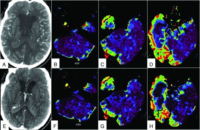
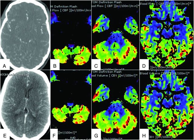
For pronouncing brain death, unlike CTP, the 2-phase CTA gives no functional information and is limited by inadvertent delay of the second acquisition, which may give false-negative results. The purpose of our study was to compare CTP and CTA derived from the CTP data with the Dupas and Frampas criteria for confirmation of brain death. A retrospective review of CTP in 11 consecutive patients for confirmation of brain death showed a sensitivity of 72.7% for 7- and 4-point scores, 81.8% for opacification of the ICV, and 100% for CTP scores in the brain stem. CTA obtained from the CTP data showed similar sensitivity in the diagnosis of brain death. This protocol also reduces the iodinated contrast dose and is less operator-dependent. The addition of the functional tools of CTP increased the sensitivity of CTA in the confirmation of brain death.
Figures
References
-
- Wijdicks EF, Varelas PN, Gronseth GS, et al. Evidence-based guideline update: determining brain death in adults—report of the Quality Standards Subcommittee of the American Academy of Neurology. Neurology 2010;74:1911–18 - PubMed
-
- Wijdicks EF. The diagnosis of brain death. N Engl J Med 2001;344:1215–21 - PubMed
-
- Wijdicks EF. Brain death worldwide: accepted fact but no global consensus in diagnostic criteria. Neurology 2002;58:20–25 - PubMed
-
- Decree No. 96–1401 of December 2, 1996 concerning brain death diagnosis, organ, tissue, and cell harvesting for therapeutic or scientific uses and Circular D.G.S No. 96–733 of December 4, 1996. Official Journal of the French Republic; 1996