. 2013 Mar 1;304(5):L350-60.
doi: 10.1152/ajplung.00243.2012. Epub 2012 Dec 28.

Metallothionein-induced zinc partitioning exacerbates hyperoxic acute lung injury

Affiliations

Metallothionein-induced zinc partitioning exacerbates hyperoxic acute lung injury

Sang-Min Lee et al. Am J Physiol Lung Cell Mol Physiol. .

Abstract

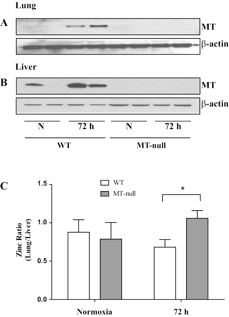
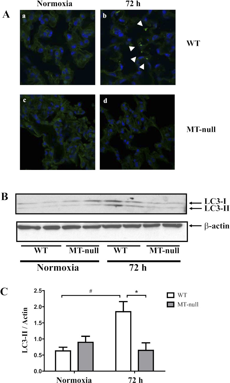
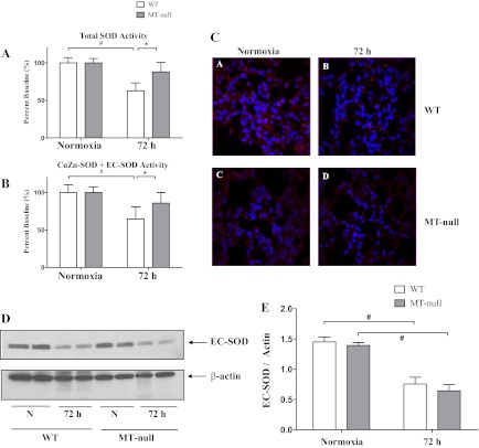
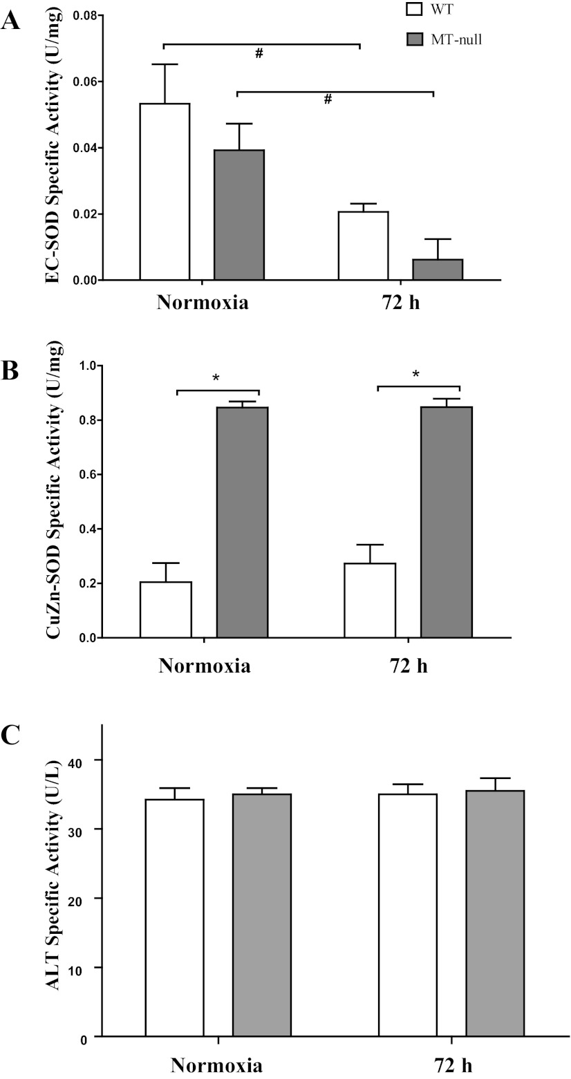
Hypozincemia, with hepatic zinc accumulation at the expense of other organs, occurs in infection, inflammation, and aseptic lung injury. Mechanisms underlying zinc partitioning or its impact on extrahepatic organs are unclear. Here we show that the major zinc-binding protein, metallothionein (MT), is critical for zinc transmigration from lung to liver during hyperoxia and preservation of intrapulmonary zinc during hyperoxia is associated with an injury-resistant phenotype in MT-null mice. Particularly, lung-to-liver zinc ratios decreased in wild-type (WT) and increased significantly in MT-null mice breathing 95% oxygen for 72 h. Compared with female adult WT mice, MT-null mice were significantly protected against hyperoxic lung injury indicated by reduced inflammation and interstitial edema, fewer necrotic changes to distal airway epithelium, and sustained lung function at 72 h hyperoxia. Lungs of MT-null mice showed decreased levels of immunoreactive LC3, an autophagy marker, compared with WT mice. Analysis of superoxide dismutase (SOD) activity in the lungs revealed similar levels of manganese-SOD activity between strains under normoxia and hyperoxia. Lung extracellular SOD activity decreased significantly in both strains at 72 h of hyperoxia, although there was no difference between strains. Copper-zinc-SOD activity was ~4× higher under normoxic conditions in MT-null compared with WT mice but was not affected in either group by hyperoxia. Collectively the data suggest that genetic deletion of MT-I/II in mice is associated with compensatory increase in copper-zinc-SOD activity, prevention of hyperoxia-induced zinc transmigration from lung to liver, and hyperoxia-resistant phenotype strongly associated with differences in zinc homeostasis during hyperoxic acute lung injury.


Figures

Fig. 1.
Fig. 1.
Metallothionein (MT) is upregulated in liver and lung tissues after hyperoxia (A and B). Three-day hyperoxia exposure increased lung (A) and liver (B) expression of MT in wild-type mice, whereas the MT-null mice had no expression of MT as expected. The loading control was β-actin, showing that lanes were similarly loaded. Representative protein bands in duplicate are presented (n = 8). In hyperoxia, zinc is partitioned into the liver (C). The lung and liver zinc contents were measured and presented as a ratio of lung-to-liver zinc distribution. At normoxic baseline (N), both wild-type (n = 8) and MT-null (n = 6) mice had similar zinc distribution between the two organs. On the other hand, following 72 h of hyperoxia (72 h), the wild-type mice (n = 11) increased the liver content of MT (as presented in Fig. 1), leading to an increased liver accumulation of zinc with subsequent lowering of lung-to-liver zinc ratio compared with the MT-null mice (n = 8). Values are means ± SD and the difference between the wild-type and MT-null mice was significant at 72 h of hyperoxia (*P = 0.02).
Fig. 2.
Fig. 2.
Both wild-type (WT) and MT-null mice had similar weight loss following hyperoxia exposure, but the WT mice had more pronounced lung injury. A: both WT and MT-null mice lost weight, and the weight loss was statistically significant as early as 48 h, but there was no difference in the rate of weight loss between WT and MT-null mice (#P < 0.05). At baseline both WT (n = 10) and MT-null (n = 6) mice had similar total cell counts in the bronchoalveolar lavage (B) and percent neutrophils (C). Although the total cell count did not change between the WT (n = 6) and MT-null (n = 6) mice by 48 h of hyperoxia, the differential count of neutrophils was significantly higher in WT mice compared with MT-null mice (*P < 0.05). The WT bronchoalveolar lavage (BAL) neutrophils were also significantly elevated at 48 h of hyperoxia compared with baseline normoxia (#P < 0.05). A similar pattern for total cell counts and percent neutrophils was observed at 72 h of hyperoxia between the WT (n = 6) and MT-null (n = 8) (*P < 0.05) as well as within the WT and MT-null groups between the baseline normoxia and 72-h time points (#P < 0.05). The lung injury was also corroborated by the measurement of BAL protein concentration (D). At 48 and 72 h, the WT had significantly higher BAL protein concentrations than the MT-null mice (*P < 0.05). At 72 h, the BAL protein concentration was also significantly elevated within the groups (#P < 0.05).
Fig. 3.
Fig. 3.
WT had more pronounced lung injury shown with the physiological measurements compared with the MT-null mice. Pulmonary mechanical function was assessed in cohorts of mice at 48 and 72 h of hyperoxia. A: hyperoxia exposure significantly decreased static compliance (Cst) in WT (n = 8, 5, and 10 at 0, 48, and 72 h, respectively) mice but was not affected in MT-null (n = 6, 10, and 9 at 0, 48, and 72 h, respectively) mice (*P < 0.05). Over the course of the experiments, the WT mice also had a loss in static compliance compared with the baseline controls (#P < 0.05). B: hyperoxia significantly impaired lung hysteresis in WT mice and the increase in pressure-volume (P-V) loop area was significantly greater in WT than MT-null mice at 48 and 72 h hyperoxia (*P < 0.05). The WT also had impaired hysteresis over time compared with the baseline controls (#P < 0.05). Tissue damping (G; C) and elastance (H; D) were similarly significantly increased in WT mice during hyperoxia but not affected in MT-null mice. *Statistically significant difference between WT and MT-null mice; #difference over time in the same strain.
Fig. 4.
Fig. 4.
WT had more pronounced lung injury at the tissue and cellular level compared with the MT-null mice. After 72 h of hyperoxia, histopathological changes were apparent in WT mice but not MT-null mice determined by light (Ab vs. Ad) and transmission electron microscopy (Bb vs. Bd). Lung tissue of hyperoxic WT mice (n = 6) showed increased cellularity and apparent interstitial edema compared with normoxic controls as well as MT-null at ×20 magnification (n = 6) mice (Ab). At the cellular level, lungs of hyperoxic WT mice (n = 4) showed damage of both type I and type II epithelial cells including presence of immature autophagosomes and necrotic changes (black arrow) with nuclear disintegration (Bb) and tissue edema (black arrowheads), whereas ultrastructure of distal epithelium appeared well preserved at 72 h of hyperoxia in MT-null mice (n = 4) (Bd) with early autophagosomes (white arrowheads).
Fig. 5.
Fig. 5.
WT had more pronounced lung injury at the tissue and cellular level compared with the MT-null mice. At baseline, the autophagy marker LC3 staining signal showed a diffuse pattern in both normoxic WT (n = 7) and MT-null mice (n = 6) (Aa and Ac). Hyperoxia exposure resulted in a punctate staining pattern for LC3, indicating the induction of autophagy in WT mice (white arrowheads) (Ab). The LC3 staining intensity was less pronounced in the MT-null mice, suggesting that autophagy was less pronounced in hyperoxia-treated lungs of MT-null mice (Ad). The Western blot analysis supports the findings of the tissue staining findings of autophagy (8). Lung tissue conversion of LC3-I to LC3-II was significantly elevated in the WT mice (n = 6) compared with MT-null mice (n = 6, *P < 0.05) (B and C). In WT mice, LC3-II protein also increased significantly at 72 h of hyperoxia compared with the normoxic state (#P < 0.05). In contrast, there were no significant changes in LC3-II protein at 72 h of hyperoxia in MT-null mice.
Fig. 6.
Fig. 6.
WT had significant decrease in total and CuZn + extracellular superoxide dismutase (EC-SOD) activity compared with the MT-null mice. The total lung antioxidant superoxide dismutase (SOD) capacities of the WT (n = 5) and MT-null mice (n = 5) at baseline were similar, whereas the total SOD activity decreased significantly over time for WT mice (n = 5, #P < 0.05); there was also significant decrease between WT and MT-null mice (n = 5) total SOD activity at 72 h hyperoxia (A). Overall zinc-dependent SOD activity was then measured, and similar changes of SOD activity were noted to occur in response to hyperoxia with a decrease in SOD activity in WT (n = 5) and no change in MT-null mice (n = 5) (#,*P < 0.05) (B). There was similar degree of loss of EC-SOD fluorescence staining following hyperoxia in both WT and MT-null mice (C). This was corroborated with the whole lung Western blot analysis, and the decrease was statistically significant for both WT (n = 5) and MT-null (n = 5) mice (#P < 0.05) (D and E).
Fig. 7.
Fig. 7.
Both WT and MT-null mice have decreased EC-SOD activity following hyperoxia. Hyperoxia caused a decrease in EC-SOD activity (A) in both WT (n = 5) and MT-null mice (n = 5). CuZn-SOD activity was not affected by hyperoxia in either WT or MT-null mice (B). CuZn-SOD activity, however, was ∼4× greater in lungs of MT-null mice (n = 5) than WT mice (n = 6) at control and at 72 h of hyperoxia (*P < 0.05). Both WT and MT-null mice have similar serum alanine aminotransferase (ALT) specific activity at baseline and following hyperoxia. ALT activity, an indirect marker of liver injury, was not affected by hyperoxia in either WT (n = 4) or MT-null (n = 4) mice (C).

Similar articles

Cited by

References

    1. Abel J, de Ruiter N. Inhibition of hydroxyl-radical-generated DNA degradation by metallothionein. Toxicol Lett 47: 191–196, 1989 - PubMed
    1. Auten RL, O'Reilly MA, Oury TD, Nozik-Grayck E, Whorton MH. Transgenic extracellular superoxide dismutase protects postnatal alveolar epithelial proliferation and development during hyperoxia. Am J Physiol Lung Cell Mol Physiol 290: L32–L40, 2006 - PMC - PubMed
    1. Bates JH, Irvin CG. Time dependence of recruitment and derecruitment in the lung: a theoretical model. J Appl Physiol 93: 705–713, 2002 - PubMed
    1. Carlsson LM, Jonsson J, Edlund T, Marklund SL. Mice lacking extracellular superoxide dismutase are more sensitive to hyperoxia. Proc Natl Acad Sci USA 92: 6264–6268, 1995 - PMC - PubMed
    1. Cousins RJ, Liuzzi JP, Lichten LA. Mammalian zinc transport, trafficking, and signals. J Biol Chem 281: 24085–24089, 2006 - PubMed

Publication types

LinkOut - more resources