

Scheimpflug imaging criteria for identifying eyes at high risk of acute angle closure
- PMID: 23275818
- PMCID: PMC3520477
Scheimpflug imaging criteria for identifying eyes at high risk of acute angle closure
Abstract
Purpose: To compare anterior segment and ocular biometric parameters in unaffectedfellow eyes of patients with a previous attack of acute angle closure (AAC), primary angle closure suspect (PACS) eyes, and normal eyes; and to identify eyes at high risk of AAC among primary angle closure suspects.
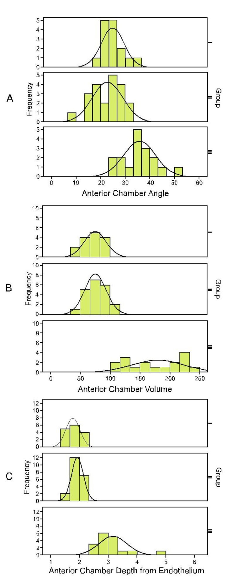
Methods: In this case-control study, 16 unaffected fellow eyes of patients with aprevious attack of AAC (group I), 20 PACS eyes (group II) and 18 normal eyes (group III) underwent Pentacam and A-scan echography.
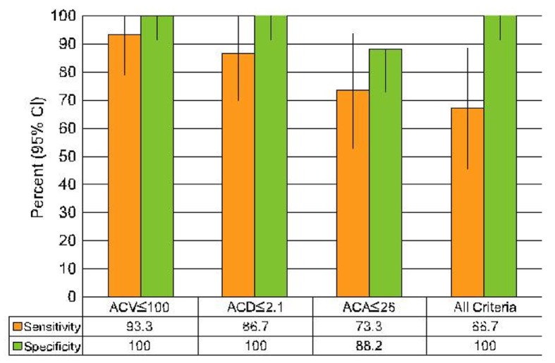
Results: Mean anterior chamber volume was 72±18, 77±18 and 176±44 µl in groupsI, II, and III, respectively (P<0.001). Corresponding values for anterior chamber angle in the same order were 24.8±4.6, 22.6±6.3 and 35.8±6.9 degrees (P<0.001), and that for anterior chamber depth measured from the corneal endothelium were 1.80±0.26, 1.93±0.23 and 3.13±0.59 mm, respectively (P<0.001). Using receiver operating characteristic (ROC) curves, anterior chamber volume ≤100 µl was associated with a high risk of AAC with sensitivity of 93.3% and specificity of 100%. Corresponding values for anterior chamber depth ≤2.1 mm were 86.7% and 100%, and that for anterior chamber angle ≤26° were 73.3% and 88.2%, respectively. Age, sex, central corneal thickness, and lens thickness were comparable among the study groups (all P values >0.05).
Conclusion: Eyes with anterior chamber volume ≤100 µl, depth ≤2.1 mm and angle≤26° may be considered at high risk for developing AAC. These criteria could be helpful for making decisions regarding prophylactic laser peripheral iridotomy.
Keywords: Acute Angle Closure; Glaucoma; Scheimpflug Imaging.
Figures
References
LinkOut - more resources
Full Text Sources