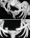
Bilateral asymmetrical traumatic sternoclavicular joint dislocations
- PMID: 23275851
- PMCID: PMC3524004
- DOI: 10.12816/0003180
Bilateral asymmetrical traumatic sternoclavicular joint dislocations
Abstract
Unilateral and bilateral sternoclavicular joint (SCJ) dislocations are rare injuries. The difficulty in assessing this condition often leads to delay in diagnosis and treatment. We report a rare case of bilateral asymmetrical traumatic SCJ dislocations in a 45-year-old male. The right anterior SCJ dislocation was reduced in the emergency room (ER) and resulted in residual instability. The left posterior SCJ dislocation was asymptomatic and unnoticed for six months. It is important for ER physicians and orthopaedic surgeons to be able identify and treat this condition. All suspected SCJ dislocations should be evaluated by computed tomography (CT) scan for confirmation of the diagnosis and evaluation of both SCJs. Posterior SCJ dislocation is a potentially fatal injury and should not be overlooked due to the presence of other injuries. Surgical intervention is often necessary in acute and old cases.
Keywords: Case report; Dislocations; Emergency; Saudi Arabia; Shoulder; Sternoclavicular joint.
Figures


References
-
- Renfree KJ, Wright TW. Anatomy and biomechanics of the acromioclavicular and sternoclavicular joints. Clin Sports Med. 2003;22:219–37. - PubMed
-
- Nettles JL, Linscheid R. Sternoclavicular dislocations. J Trauma. 1968;8:158–64. - PubMed
-
- Camara ES, Bousso A, Tall M, Sy MH. Posterior sternoclavicular dislocations. Eur J Orthop Surg Traumatol. 2009;19:7–9.
-
- Chien LC, Hsu IL, Tsai MC, Lo CJ. Bilateral anterior sternoclavicular dislocation. J Trauma. 2009;66:1504. - PubMed
-
- Yeh GL, Williams GR., Jr Conservative management of sternoclavicular injuries. Orthop Clin North Am. 2000;31:189–203. - PubMed
LinkOut - more resources
Full Text Sources
