. 2012 Dec;2(12):1687-701.
doi: 10.1534/g3.112.003814. Epub 2012 Dec 1.

Suppressors of ipl1-2 in components of a Glc7 phosphatase complex, Cdc48 AAA ATPase, TORC1, and the kinetochore

Affiliations

Suppressors of ipl1-2 in components of a Glc7 phosphatase complex, Cdc48 AAA ATPase, TORC1, and the kinetochore

Lucy C Robinson et al. G3 (Bethesda). 2012 Dec.

Abstract

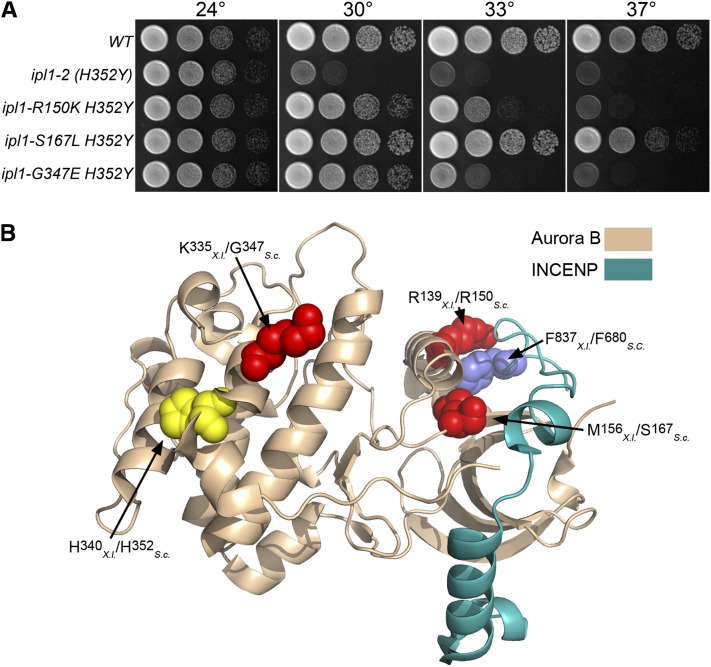
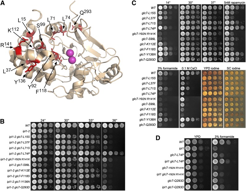
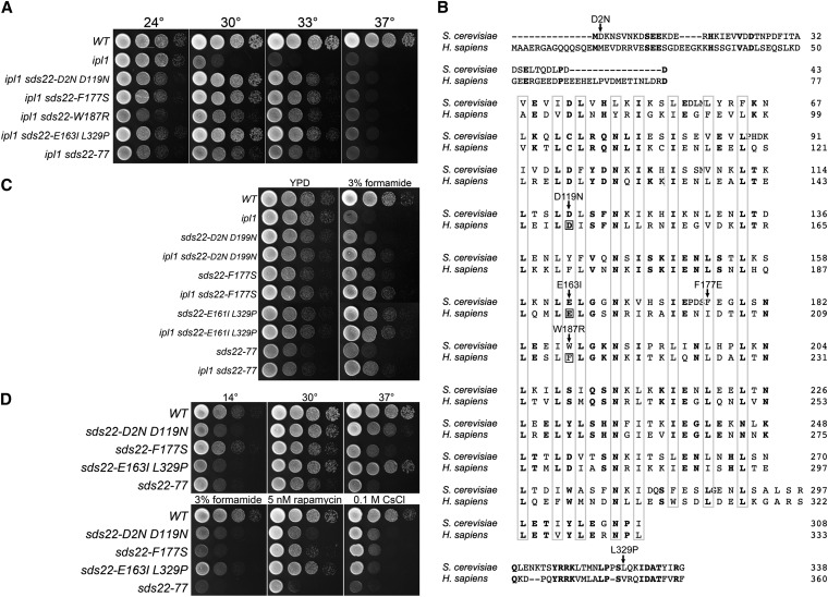
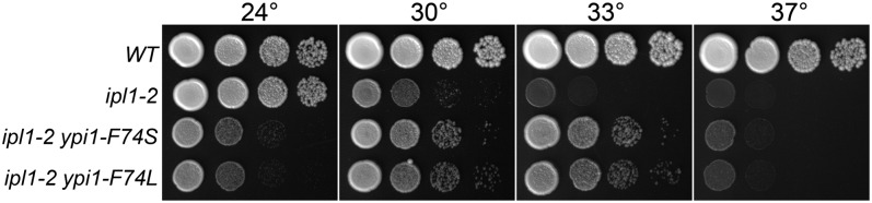
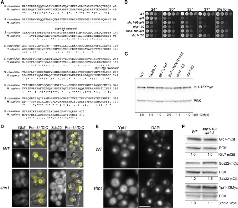
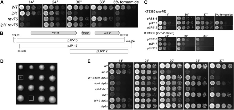
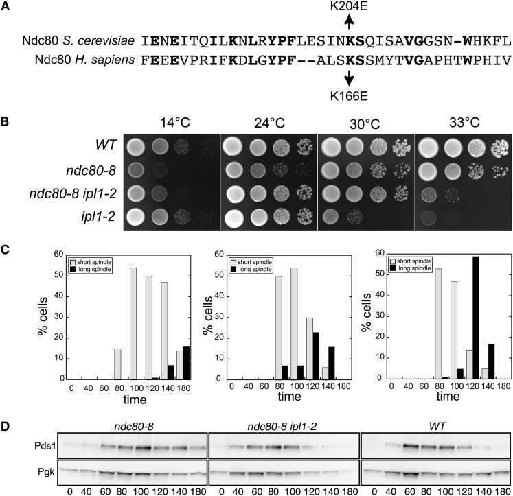
Ipl1/Aurora B is the catalytic subunit of a protein kinase complex required for chromosome segregation and nuclear division. Before anaphase, Ipl1 is required to establish proper kinetochore-microtubule associations and to regulate the spindle assembly checkpoint (SAC). The phosphatase Glc7/PP1 opposes Ipl1 for these activities. To investigate Ipl1 and Glc7 regulation in more detail, we isolated and characterized mutations in the yeast Saccharomyces cerevisiae that raise the restrictive temperature of the ipl-2 mutant. These suppressors include three intragenic, second-site revertants in IPL1; 17 mutations in Glc7 phosphatase components (GLC7, SDS22, YPI1); two mutations in SHP1, encoding a regulator of the AAA ATPase Cdc48; and a mutation in TCO89, encoding a subunit of the TOR Complex 1. Two revertants contain missense mutations in microtubule binding components of the kinetochore. rev76 contains the missense mutation duo1-S115F, which alters an essential component of the DAM1/DASH complex. The mutant is cold sensitive and arrests in G2/M due to activation of the SAC. rev8 contains the missense mutation ndc80-K204E. K204 of Ndc80 corresponds to K166 of human Ndc80 and the human Ndc80 K166E variant was previously shown to be defective for microtubule binding in vitro. In a wild-type IPL1 background, ndc80-K204E cells grow slowly and the SAC is activated. The slow growth and cell cycle delay of ndc80-K204E cells are partially alleviated by the ipl1-2 mutation. These data provide biological confirmation of a biochemically based model for the effect of phosphorylation on Ndc80 function.

Keywords: Aurora B; GLC7; IPl1; NDC80; kinetochore.


Figures

Figure 1
Figure 1
Intragenic ipl1-2 suppressors. (A) Cultures of WT (KT1112), ipl1-2 (H352Y) (KT1829), ipl1-2 R150K H352Y (KT2865), ipl1-S167L H352Y (KT2867), and ipl1-G347E H352Y (KT2869) strains were serially diluted onto YPD medium and imaged after 40 hr at the designated temperatures. (B) The locations of intragenic ipl1-2 suppressor mutations are mapped on the X. laevis Aurora B- INCENP structure [2BFX.pdb (Sessa et al. 2005)]. The highlighted amino acid residues are represented as space-filling models. The location of the ipl1-2 mutation H352Y is shown in yellow. Residues altered by intragenic suppressor mutations are red, and the amino acid residue in INCENP that associates with R139 is blue. Note that both S167 and R150 are predicted to lie near the interface with INCENP/Sli15.
Figure 2
Figure 2
Extragenic ipl1-2 suppressors in GLC7. (A) Locations of GLC7 ipl1-2 suppressor changes mapped on the structure of rabbit PP1α [1FJM.pdb (Goldberg et al. 1995)]. The amino acid residues altered in the ipl1 suppressor mutants are stick representations in red. The two Mn2+ ions in the active site are in magenta. (B) Cultures of WT (KT1113), ipl1-2 (KT1829), ipl1-2 glc7-L15S (KT3062), ipl1-2 glc7-L37F (KT2940), ipl1-2 glc7-L71S (KT2938), ipl1-2 glc7-L74P (KT3361), ipl1-2 glc7-Y92N R141K (KT3365), ipl1-2 glc7-S99L (KT2939), ipl1-2 glc7-K112E (KT3064), ipl1-2 glc7-F118S (KT3059), ipl1-2 glc7-Y136N (KT3358), and ipl1-2 glc7-Q293D (KT3066) strains were serially diluted onto YPD medium and imaged after 40 hr at the designated temperatures. (C) Cultures of WT (KT1113), glc7-L15S (KT3304), glc7-L37F (KT2973), glc7-L71S (KT2969), glc7-L74P (KT3359), glc7-Y92N R141K (KT3363), glc7-S99L (KT2970), glc7-K112E (KT3308), glc7-F118S (KT3302), glc7-Y136N (KT3355), and glc7-Q293D (KT3310) strains were serially diluted onto the designated medium and imaged at the designated temperatures. All media, with the exception of SC, are either YPD or YPD supplemented with the indicated components. The YPD 14° plates were incubated for 14 d. All other plates were incubated for 40 hr. YPD iodine and SC iodine panels indicate cells grown on YPD or SC media for 40 hr and then stained with iodine vapor. (D) Cultures of WT (KT1113), ipl1-2 (KT1829), glc7-L74P (KT3359), ipl1-2 glc7-L74P (KT3361), glc7-Y92N R141K (KT3363), ipl1-2 glc7-Y92N R141K (KT3365), glc7-Q293D (KT3310), and ipl1-2 glc7-Q293D (KT3066) strains were serially diluted onto YPD medium and YPD medium containing 3% formamide and imaged after 24 and 72hr, respectively, at 24°.
Figure 3
Figure 3
Extragenic ipl1-2 suppressors in SDS22. (A) Cultures of WT (KT1113), ipl1-2 (KT1829), ipl1-2 sds22-D2N D119N (KT2934), ipl1-2 sds22-F177S (KT3353), ipl1-2 sds22-W187R (KT3381), ipl1-2 sds22-E163I L329P (KT3052), and ipl1-2 sds22-77 (KT2936) strains were serially diluted onto YPD medium and imaged after 40 hr at the designated temperatures. (B) ClustalW alignment of human and S. cerevisiae Sds22 proteins showing locations of ipl1 suppressor mutations. Boxed residues correspond to sites of mutations in human Sds22 that prevent proper binding of PP1 (Ceulemans et al. 2002). (C) Cultures of WT (KT1113), ipl1-2 (KT1829), sds22-D2N D119N (KT2963), ipl1-2 sds22-D2N D119N (KT2934), sds22-F177S (KT3351), ipl1-2 F177S (KT3353), sds22-E163I L329P (KT3292), ipl1-2 sds22-E163I L329P (KT3052), sds22-77 (KT2964), and ipl1-2 sds22-77 (KT2936) strains were serially diluted onto YPD medium and YPD medium containing 3% formamide and imaged after 24 hr and 72hr at 24°, respectively. (D) Cultures of WT (KT1113), sds22-D2N D119N (KT2963), sds22-F177S (KT3351), sds22-E163I L329P (KT3292), and sds22-77 (KT2964) strains were serially diluted onto the designated medium at the designated temperatures. Media, with the exception of SC, are either YPD or YPD supplemented as indicated. The YPD 14° plates were incubated for 14 d. All other plates were incubated for 40 hr.
Figure 4
Figure 4
Extragenic ipl1-2 suppressors in YPI1. Cultures of WT (KT1113), ipl1-2 (KT1829), ipl1-2 ypi1-F74S (KT3368), and ipl1-2 ypi1-F74L (KT3370) strains were serially diluted onto YPD medium and imaged after 40 hr at the designated temperatures.
Figure 5
Figure 5
Extragenic ipl1-2 suppressors in SHP1. (A) Sequence alignment of human p47 and S. cerevisiae Shp1 proteins with the locations of ipl1 suppressor mutations indicated. (B) Cultures of WT (KT1113), ipl1-2 (KT1829), ipl1-2 shp1-99 (KT3413), shp1-99 (KT3412), ipl1-2 shp1-105 (KT3419), and shp1-105 (KT3416) strains were serially diluted onto YPD medium and imaged after 40 hr at the designated temperatures. (C) Immunoblot analysis of extracts from WT (KT3383), tco89-71 (KT3389), glc7-L74P (KT3391), glc7-Y92N R141K (KT3392), shp1-105 (KT105), and shp1-99 (KT3396) strains containing IPL1-13Myc. Ipl1-13Myc levels indicated under the lanes were calculated relative to the loading control (Pgk1) signal and normalized to the WT. (D) Fluorescence microscopy of Glc7-mCitrine and Sds22-mCitrine in WT (KT3242 and KT2856) and shp1-105 (KT3424 and KT3428) cells grown to log phase in YPD medium at 24°. The right panels of each set are overlays of Pom34-mCherry and DIC images to demarcate the nuclear periphery. Note that fluorescence images for the Glc7-mCitrine and Sds22-mCitrine are normalized independently. The actual fluorescence levels are much greater in the Glc7-mCitrine strains. Scale bar: 5 μm. (E) Indirect immunofluorescence of Ypi1-13Myc in WT (KT2881) and shp1-105 (KT3427) cells. Scale bar: 5 μm. (F) Immunoblot analysis of extracts from WT and shp1-105 ipl1-2 strains containing Glc7-mCitrine (KT3242 and KT3424), Sds22-mCitrine (KT2856 and KT3428), or Ypi1-13Myc (KT2881 and KT3427). Protein levels were calculated relative to the signal in the loading control (Pgk1) and normalized to the WT.
Figure 6
Figure 6
Extragenic ipl1-2 suppressors in DUO1. (A) Cultures of WT (KT1113), ipl-2 (KT1829), rev76 (KT3385), and ipl1-2 rev76 (KT3386) strains were serially diluted onto YPD medium and imaged after 40 hr at the indicated temperatures. (B) Genetic map of the PYC1-DUO1-YBP2 region of chromosome VII showing the locations of genomic DNA fragments that complement the rev76 suppressor phenotype. (C) Cultures of duo1-S115F (KT3385) and ipl1-2 duo1-S115F (KT3386) mutant strains transformed with the designated plasmids were serially diluted onto the following media: Top panels: YPD at the designated temperatures and YPD + 3% formamide at 30°. Bottom panels: Synthetic medium lacking uracil at the designated temperatures. (D) Images of tetrads from a cross between duo1-S115F and mad1::HIS3 strains (KT3386 X KT1688). The boxes identify the duo1-S115F mad1::HIS3 double mutants. These were the largest double mutant colonies observed; the majority of double mutants failed to grow into macroscopic colonies. Each column represents the four spore clones of a tetrad. (E) Cultures of WT (KT1113), ipl-2 (KT1829), ipl1-2 duo1-S115F ybp2Δ::kanMX6 (KT3409), duo1-S115F ybp2Δ::kanMX6 (KT3410), ipl1-2 duo1-S115F (KT3386), duo1-S115F (KT3385), ipl1-2 ybp2Δ::kanMX6 (KT3401), and ybp2Δ::kanMX6 (KT3403) strains were serially diluted onto YPD medium and imaged after 40 hr at the indicated temperatures with the exception of the plate at 14°, which was imaged after 7 d.
Figure 7
Figure 7
Extragenic ipl1-2 suppressors in NDC80. (A) Sequence alignment of yeast and human Ndc80 proteins. (B) Cultures of WT (KT1113), ndc80-K204E (ndc80-8) (KT3255), ndc80-K204E ipl1-2 (KT3257), and ipl1-2 (KT1963) strains were serially diluted, plated onto indicated media, and imaged after 44 hr. The first four panels show YPD medium incubated at the designated temperatures. (C) and (D) Cultures of ndc80-K204E(KT3317), ndc80-K204E ipl1-2 (KT3320), and WT (KT3319) cells were arrested with alpha factor, released into YPD medium at 24°, and monitored for spindle length and entrance into anaphase using Spc42-3XGFP and Pds1-13Myc fusions, respectively, at the designated time points. (C) Quantitation of the percentage of cells with short (≤2.0 μm) or long (>2.0 μm) spindles. At least 64 mitotic spindles were measured at each time point for each strain. (D) Immunoblot analysis of Pds1-13XMyc in whole cell extracts. Pgk serves as the loading control.

Similar articles

Cited by

References

    1. Akiyoshi B., Nelson C. R., Ranish J. A., Biggins S., 2009a Analysis of Ipl1-mediated phosphorylation of the Ndc80 kinetochore protein in Saccharomyces cerevisiae. Genetics 183: 1591–1595 - PMC - PubMed
    1. Akiyoshi B., Nelson C. R., Ranish J. A., Biggins S., 2009b Quantitative proteomic analysis of purified yeast kinetochores identifies a PP1 regulatory subunit. Genes Dev. 23: 2887–2899 - PMC - PubMed
    1. Altschul S. F., Gish W., Miller W., Myers E. W., Lipman D. J., 1990. Basic local alignment search tool. J. Mol. Biol. 215: 403–410 - PubMed
    1. Andrews P. D., Stark M. J., 2000. Dynamic, Rho1p-dependent localization of Pkc1p to sites of polarized growth. J. Cell Sci. 113: 2685–2693 - PubMed
    1. Andrews P. D., Ovechkina Y., Morrice N., Wagenbach M., Duncan K., et al. , 2004. Aurora B regulates MCAK at the mitotic centromere. Dev. Cell 6: 253–268 - PubMed

Publication types

MeSH terms

LinkOut - more resources