. 2012 Oct 19;2(4):112.
doi: 10.4172/2165-7491.1000112.

Nuclear routing networks span between nuclear pore complexes and genomic DNA to guide nucleoplasmic trafficking of biomolecules

Affiliations

Nuclear routing networks span between nuclear pore complexes and genomic DNA to guide nucleoplasmic trafficking of biomolecules

Marek Malecki et al. J Fertili In Vitro. .

Abstract

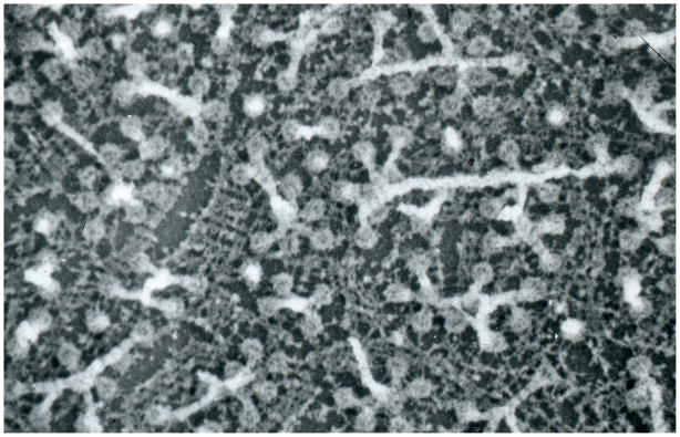
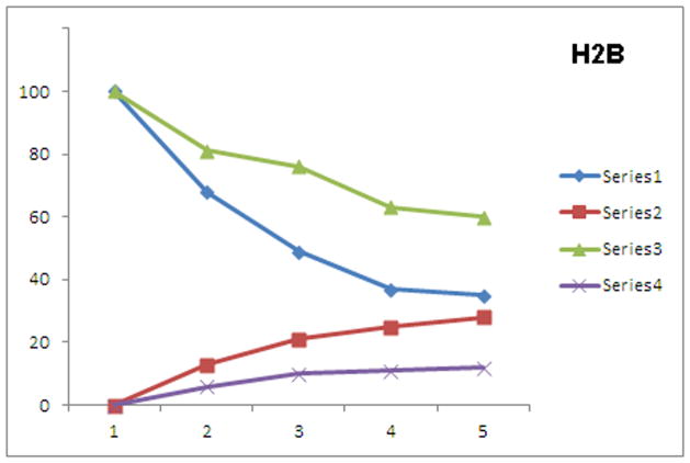
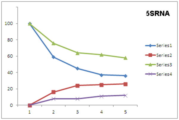
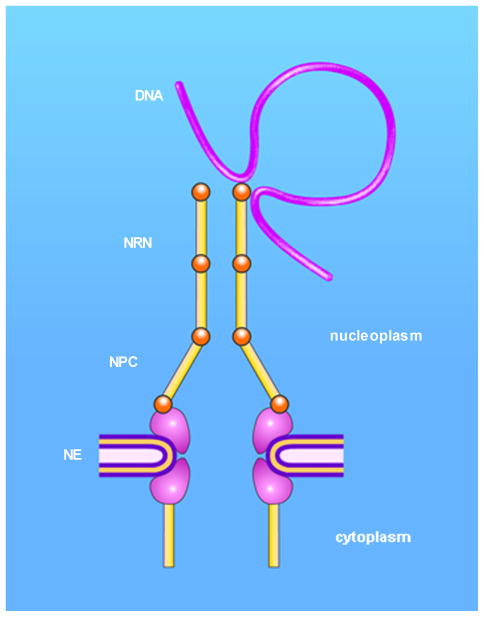
In health and disease, biomolecules, which are involved in gene expression, recombination, or reprogramming have to traffic through the nucleoplasm, between nuclear pore complexes (NPCs) and genomic DNA (gDNA). This trafficking is guided by the recently revealed nuclear routing networks (NRNs).In this study, we aimed to investigate, if the NRNs have established associations with the genomic DNA in situ and if the NRNs have capabilities to bind the DNA de novo. Moreover, we aimed to study further, if nucleoplasmic trafficking of the histones, rRNA, and transgenes' vectors, between the NPCs and gDNA, is guided by the NRNs.We used Xenopus laevis oocytes as the model system. We engineered the transgenes' DNA vectors equipped with the SV40 LTA nuclear localization signals (NLS) and/or HIV Rev nuclear export signals (NES). We purified histones, 5S rRNA, and gDNA. We rendered all these molecules superparamagnetic and fluorescent for detection with nuclear magnetic resonance (NMR), total reflection x-ray fluorescence (TXRF), energy dispersive x-ray spectroscopy (EDXS), and electron energy loss spectroscopy (EELS).The NRNs span between the NPCs and genomic DNA. They form firm bonds with the gDNA in situ. After complete digestion of the nucleic acids with the RNases and DNases, the newly added DNA - modified with the dNTP analogs, bonds firmly to the NRNs. Moreover, the NRNs guide the trafficking of the DNA transgenes' vectors - modified with the SV40 LTA NLS, following their import into the nuclei through the NPCs. The pathway is identical to that of histones. The NRNs also guide the trafficking of the DNA transgenes' vectors, modified with the HIV Rev NES, to the NPCs, followed by their export out of the nuclei. Ribosomal RNAs follow the same pathway.To summarize, the NRNs are the structures connecting the NPCs and the gDNA. They guide the trafficking of the biomolecules between the NPCs and the gDNA.


Conflict of interest statement

CONFLICT OF INTEREST

The authors declare no conflict of interest. The authors own the IP pertinent to this work, while protected at the WIPO and USPTO.

Figures

Fig. 1
Fig. 1. Work-flow of the project
The Xenopus laevis oocytes were microinjected into the cytoplasm or nucleoplasm with the transgenes’ vectors or biomolecules (a). The nuclei were isolated (b). The isolated nuclei were, either cryo-immobilized and homogenized (c), or attached to the sticky glass (d). The gDNA, RNA, and proteins were isolated from the homogenized nuclei for NMRS and TRXFS. The nuclear envelopes architecture, including lamins, NPCs, and NRNs were exposed after rinsing off nucleoplasm, digestion with DNases and RNases. The DNA binding assays were performed with the EDXS and TRXFS.
Fig. 2
Fig. 2. Architecture of the nuclear routing networks (NRNs)
The nucleoplasm was removed to expose the architecture of the ultrastructures assembled onto the nucleoplasmic side of the nuclear envelope. The most prominent are the long stretches of the nuclear routing networks (NRNs). The NRNs project from the tops of the nuclear pore complexes (NPCs) inside of the nucleus. The bases of the NPCs are inserted into the lamin network of the nuclear envelope. Horizontal field width (HFW): 6960nm.
Fig. 3
Fig. 3. Ultrastructure of the nuclear routing networks (NRNs)
The nucleoplasm was removed followed by cryoimmobilization, freeze-substitution, embedding, and cutting 250 nm thick sections perpendicular to the nuclear envelope. The sections were imaged with the electron energy loss spectroscopy (EELS) at the zero energy loss and contrast tuning. This thick sections contained the entire NPCs and long portions of the NPC (these structures could not be contained within 50 nm thin sections for TEM), but resulted in some overlap of structures (they can be resolved on the stereo pairs or with the movies from 3D reconstructions). The long stretches of the nuclear routing networks (NRNs) project from the nuclear envelope (upwards). HFW: 840 nm.
Fig. 4
Fig. 4. Associations of the gDNA with the NRNs
The nucleoplasm was removed to expose structures of the nuclear envelopes. Thereafter, the samples were labeled with the Fvs targeting genomic DNA, while chelating Gd. The architecture of the NRN was revealed with the secondary emission (Fig. 4a). The distribution of the gDNA attached to the NRNs was revealed in the EDXS by the elemental mapping of Gd (Fig. 4b). An overlay of 4b over 4a reveals that the gDNA is firmly anchored to the NRNs (Fig. 4c). Horizontal field width (HFW): 1100nm.
Fig. 5
Fig. 5. Statistical analysis of associations of the gDNA with the NRNs
The NRN preparations were labeled as in the fig. 4. Scintillation counts were acquired from ten randomly selected areas of ten different samples. The samples which were not labeled, labeled with the tag-free Fvs prior to the tagged Fv, and labeled with the isotype Fv were measured as the controls. The statistical significance of the counts was calculated with the t test and approved with the P value < 0.0001.
Fig. 6
Fig. 6. DNA binding assay
The nuclei were treated with DNases and RNases followed by thorough rinsing and exposure to the modified DNA tagged with Eu. Thereafter, the samples were cryoimmobilized, freeze-dried, and cryo-coated. The overview of the nucleoplasmic side of the nuclear envelope is shown with the secondary emission (SE) (Fig. 6a). The topography of the modified DNA bonded to the NRNs is shown with the binary map of Eu distribution in EDXS (Fig. 6b). An overlay of 6b over 6a reveals that the gDNA is firmly anchored to the NRNs (Fig. 6c). Horizontal field width 960 nm.
Fig. 7
Fig. 7. Statistical analysis of the DNA binding assay
The NRN samples were prepared as in the fig. 6. Scintillation counts were acquired from ten randomly selected areas of ten different samples. The samples, which were entirely depleted of the nucleic acids, labeled with the tag-free DNA prior to the binding the DNA modified with the chelated dNTP analogs, and labeled with the mixed tagged and non-tagged DNA were measured as the controls. The statistical significance of the counts was calculated with the t test and approved with the P value < 0.0001.
Fig. 8
Fig. 8. Trafficking of the SV40 LTA NLS-modified transgenes’ vectors
Quantification of trafficking of the transgenes’ vectors with (Series 1 and 2) and without (Series 3 and 4) the SV40 LTA NLS was determined after the oocytes were microinjected into the perinuclear space and rapidly cryoimmobilized at the 45 min intervals. Since the transgenes’ vectors carried Gd, then they were easily quantified with the x-ray emission. The scintillation counts were allocated to the cell compartments from the ten read-outs, averaged, normalized at each time point, and presented as the graph of the relative concentrations in the compartments. The labeling on the graph: NLS carrying vectors in the perinucler compartment (blue diamonds) and in the NRNs (red squares); the no-NLS vectors in the perinuclear compartment (green triangles) and in the NRNs (yellow Xs). The concentrations of the NLS modified vectors was rapidly falling in the perinuclear compartments and rising in the NRNs, which indicated the vectors entry and trafficking in the NRNs. Absence of the NLS resulted in retention of the vectors in the perinuclear volume and their absence in the NRNs. For clarity of the graphs only the means values were displayed. The start- and end-point values were statistically analyzed and approved with the statistical significance from the t test at the P value < 0.05.
Fig. 9
Fig. 9. Trafficking of the histones
Quantification of trafficking of the histones was determined the same way as in the figure 8. The labeling on the graph: histones in the perinucler compartment (blue diamonds), in the NRNs (red squares), intranuclear competition assay by co-microinjecting the equimolar non-chelating histones in the perinuclear compartment (green triangles) and in the NRNs (yellow Xs). The histones’ concentration in the perinuclear area was rapidly falling, while in the NRNs was rapidly increasing. Co-microinjection of the non-chelating histones resulted in the competitive effects. The start- and end-point values were statistically analyzed and approved with the statistical significance from the t test at the P value < 0.05.
Fig. 10
Fig. 10. Distribution of the HIV Rev NES-modified transgenes’ vectors
Quantification of trafficking of the transgenes’ vectors with (Series 1 and 2) and without (Series 3 and 4) the HIV Rev NES was determined after the intranuclear microinjections of the oocytes followed by rapid cryoimmobilization at the 45 min intervals. Since the transgenes’ vectors carried Eu, then they were easily quantified with the x-ray emission. The labeling on the graph: NES-modified vectors in the nuclear compartment (blue diamonds) and in the NRNs (red squares); the no-NES vectors in the nuclear compartment (green triangles) and in the NRNs (yellow Xs). The concentration of the NES-modified vectors in the nucleoplasm was rapidly falling after microinjections. The concentration of the vectors in the NRNs fractions rapidly increased, what was indicative of the entry in and passage through the NRNs. The concentrations of the transgenes’ vectors were constantly increasing outside of the nuclei as the result of the constant export of the vectors driven by the NES. Absence of the NES resulted in the retention in the nucleoplasm and not entering the NRNs. The start- and endpoint values were statistically analyzed and approved with the statistical significance from the t test at the P value < 0.05.
Fig. 11
Fig. 11. Trafficking of the rRNA
Quantification of trafficking of the 5S RNA modified with chelated Tb (Series 1 and 2) and competitive with non-chelating 5S RNA (Series 3 and 4) was determined after the intranuclear microinjections as described in the figure 10. The labeling on the graph: 5S RNA in the nuclear compartment (blue diamonds) and in the NRNs (red squares); competitive with non-chelating 5S RNA in the nuclear compartment (green triangles) and in the NRNs (yellow Xs). The concentration of the modified rRNA in the nucleoplasm was rapidly falling after microinjections. The concentration in the NRNs fractions showed the steady increase. The concentrations were constantly increasing outside of the nuclei as the result of the constant export. Co-microinjection of the non-chelating 5S RNA resulted in the competitive effects, as reflected in the reduced scintillation counts. The start- and end-point values were statistically analyzed and approved with the statistical significance from the t test at the P value < 0.05.
Fig. 12
Fig. 12. The NRNs span between the NPCs and the gDNA
The NRN spans between the NPC and the gDNA. The NRN is a molecular structure supporting the trafficking mechanisms of the rRNAs and histones, as well as, the transgenes’ vectors modified with the NLS and NES. The molecular identities of the structural components and interfacing molecules have yet to be identified and incorporated into this model.

Similar articles

Cited by

References

    1. Capecchi MR. How close are we to implementing gene targeting in animals other than the mouse? Proc Natl Acad Sci U S A. 2000;97(3):956–7. - PMC - PubMed
    1. Bunting M, Bernstein KE, Greer JM, Capecchi MR, Thomas KR. Targeting genes for self-excision in the germ line. Genes Dev. 1999;13(12):1524–8. - PMC - PubMed
    1. Turan S, Bode J. Site-specific recombinases: from tag-and-target- to tag-and-exchange-based genomic modifications. FASEB J. 2011;25(12):4088–107. - PubMed
    1. Hirano N, Muroi T, Takahashi H, Haruki M. Site-specific recombinases as tools for heterologous gene integration. Appl Microbiol Biotechnol. 2011;92(2):227–39. - PubMed
    1. Xue H, Wu S, Papadeas ST, Spusta S, Swistowska AM, MacArthur CC, Mattson MP, Maragakis NJ, Capecchi MR, Rao MS, Zeng X, Liu Y. A targeted neuroglial reporter line generated by homologous recombination in human embryonic stem cells. Stem Cells. 2009;27(8):1836–46. - PMC - PubMed

LinkOut - more resources