

A rare case of a giant cavernous lymphangioma of the chest wall and axilla in an adult patient
- PMID: 23276757
- PMCID: PMC3540229
- DOI: 10.1016/j.ijscr.2012.11.009
A rare case of a giant cavernous lymphangioma of the chest wall and axilla in an adult patient
Abstract
Introduction: Lymphangiomas are benign lesions that are most commonly encountered in the neck of small infants, but are quite uncommon in the adult population. Their removal can be quite difficult, when they reach enormous dimensions or they develop in critical locations. Complete resection is curative, but incomplete resections entail the risk of relapse. Lymphangiomas of the chest wall are quite rare.
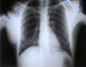
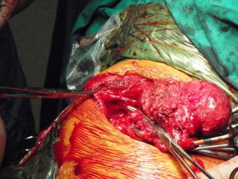
Presentation of case: We report a case of a 35-year old man with a giant cavernous lymphangioma of the right lateral chest wall extending into the axilla, which was removed en bloc.
Discussion: These tumors of lymphatic origin tend to grow, as is the case in our patient, but they also tend to recur if not resected completely. Clinical examination and chest CT scan may provide a clue to the diagnosis.
Conclusion: Lymphangiomas of the thoracic wall are extremely rare lesions and wide resection is recommended due to the high recurrence rate of these benign lesions.
Copyright © 2012 Surgical Associates Ltd. Published by Elsevier Ltd. All rights reserved.
Figures


References
-
- Ravitch M.M., Rush B.F., Jr. Cystic hygroma. In: Welch Ki, Randolph J.G., Ravitch M.M., O’Neill J.A., Jr., Rowe M.I., editors. vol. 1. Year Book Medical Publishers Inc.; Chicago: 1986. p. 536. (Paediatric surgery).
-
- Robbins S.L., Cotran R.S., Kumar V. Blood vessels. In: Robbins S.L., Cotran R.S., Kumar V., editors. Pathologic barir of dir. WB Saunders Co; Philadelphia: 1984. p. 544.http://www.jpgmonline.com/article.asp?issn=0022-3859;year=1995;volume=41...
-
- Ardenghy M., Miura Y., Kovach R., Hochberg J. Cystic hygroma of the chest wall: a rare condition. Annals of Plastic Surgery. 1996;37(2):211–213. - PubMed
-
- Fonnkaisurd E.W. Disorders of the lymphatic system. In: Welch K.J., Randolph J.G., Ravitch M.M., O’Neill J.A. Jr., Rowe M.I., editors. vol. 2. Year Book Medical Publishers Inc.; Chicago: 1986. p. 1506. (Paediatric surgery).
-
- Krainick-Strobel U., Kraimer B., Walz-Mattmuller R. Massive cavernous lymphangioma of the breast and thoracic wall: case report and literature review. Lymphology. 2006;39(September (3)):147–151. - PubMed
LinkOut - more resources
Full Text Sources
Other Literature Sources
Research Materials
