Review
. 2013 Jan 1;18(1):312-23.
doi: 10.2741/4102.

A proposal: Source of single strand DNA that elicits the SOS response

Affiliations
Review

A proposal: Source of single strand DNA that elicits the SOS response

Chiara Indiani et al. Front Biosci (Landmark Ed). .

Abstract

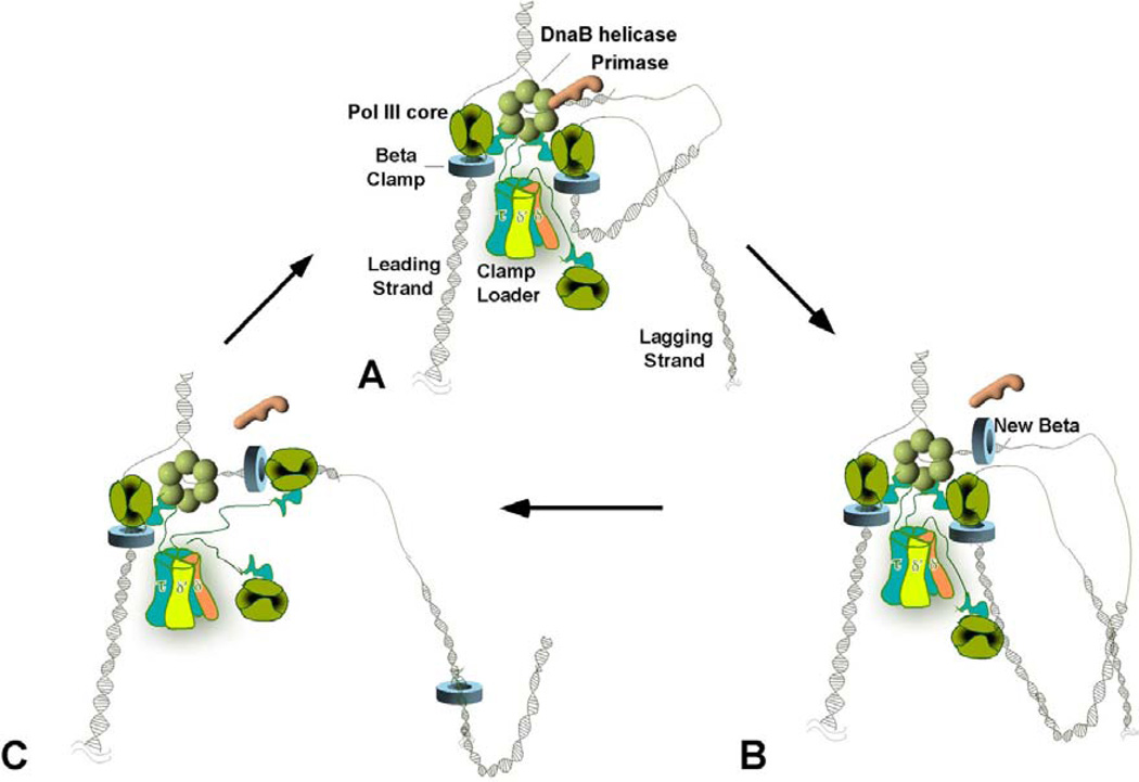
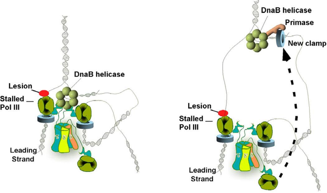
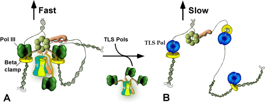
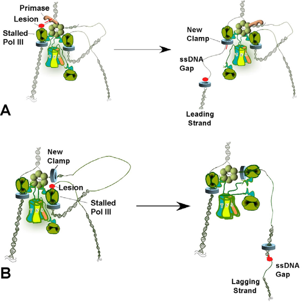
Chromosome replication is performed by numerous proteins that function together as a "replisome". The replisome machinery duplicates both strands of the parental DNA simultaneously. Upon DNA damage to the cell, replisome action produces single-strand DNA to which RecA binds, enabling its activity in cleaving the LexA repressor and thus inducing the SOS response. How single-strand DNA is produced by a replisome acting on damaged DNA is not clear. For many years it has been assumed the single-strand DNA is generated by the replicative helicase, which continues unwinding DNA even after DNA polymerase stalls at a template lesion. Recent studies indicate another source of the single-strand DNA, resulting from an inherently dynamic replisome that may hop over template lesions on both leading and lagging strands, thereby leaving single-strand gaps in the wake of the replication fork. These single-strand gaps are proposed to be the origin of the single-strand DNA that triggers the SOS response after DNA damage.


Figures

Figure 1
Figure 1
Organization of a Trimeric Replicase at the E. coli Replication Fork. The figures show a replication fork containing three Pol III core subunits bound to the same clamp loader during leading and lagging strand synthesis on an undamaged template. In (A), two Pol III cores (dark green) function on the lagging strand, one of which is illustrated off DNA. The tau subunits (blue) of the clamp loader are represented with a flexible linker that connects the clamp loader to the DnaB helicase (light green hexamer) and Pol III core. (B) After synthesis of the RNA primer, the clamp loader displaces the primase (orange) and loads the clamp onto the new primer/template junction. The two lagging-strand Pol III cores are depicted here as extending two Okazaki fragments at the same time, producing two lagging-strand loops. (C) After an Okazaki fragment is fully extended the lagging strand polymerase recycles to the newly loaded clamp and starts elongation of a new fragment, leaving the old clamp behind. This completes a full cycle of lagging strand synthesis. Fork unwinding and leading strand synthesis continue throughout the cycle.
Figure 2
Figure 2
Runaway helicase model of how ssDNA is generated during DNA damage. The leading strand polymerase stalls upon encountering a lesion (red circle) (Left). Despite a block in the leading strand, lagging-strand synthesis proceeds, implying transient uncoupling of concurrent leading/lagging strand synthesis. This creates a long ssDNA gap on the leading strand template (Right).
Figure 3
Figure 3
Pol III and TLS replisomes. Conversion of the coupled Pol III replisome to an uncoupled alternative TLS replisome. A) The coupled trimeric Pol III holoenzyme-DnaB replisome. B) Take-over of beta clamps by a TLS Pol displaces Pol III form the fork, resulting in a TLS Pol-replisome in which TLS Pols act distributively on beta clamps.
Figure 4
Figure 4
Lesion skipping model of ssDNA generation during DNA damage. (A) Leading strand lesion. The leading strand polymerase stalls upon encountering a lesion (left). The helicase recruits primase to reinitiate leading strand synthesis ahead of the lesion, leaving a single-strand gap (right). If stalling causes the replication fork to collapse, additional factors (e.g., PriA or PriC) are required to reload the helicase at the collapsed fork. (B) Lagging strand lesion. Upon encountering a lesion on the lagging strand template (Left), leading strand synthesis continues and the stalled lagging strand polymerase recycles to a new primer/template junction, leaving a single-strand gap with a template lesion (right).

References

    1. Courcelle J, Khodursky A, Peter B, Brown PO, Hanawalt PC. Comparative gene expression profiles following UV exposure in wild-type and SOS-deficient Escherichia coli. Genetics. 2001;158(1):41–64. - PMC - PubMed
    1. Walker GC. Mutagenesis and inducible responses to deoxyribonucleic acid damage in Escherichia coli. Microbiol Rev. 1984;48(1):60–93. - PMC - PubMed
    1. Fernandez De Henestrosa AR, Ogi T, Aoyagi S, Chafin D, Hayes JJ, Ohmori H, Woodgate R. Identification of additional genes belonging to the LexA regulon in Escherichia coli. Mol Microbiol. 2000;35(6):1560–1572. - PubMed
    1. Janion C. Inducible SOS response system of DNA repair and mutagenesis in Escherichia coli. Int J Biol Sci. 2008;4(6):338–344. - PMC - PubMed
    1. Little JW. Autodigestion of lexA and phage lambda repressors. Proc Natl Acad Sci U S A. 1984;81(5):1375–1379. - PMC - PubMed

Publication types

LinkOut - more resources