Review
. 2013 Jan 1;5(1):134-48.
doi: 10.2741/s362.

Protective cardiovascular and renal actions of vitamin D and estrogen

Affiliations
Review

Protective cardiovascular and renal actions of vitamin D and estrogen

Pandu R Gangula et al. Front Biosci (Schol Ed). .

Abstract

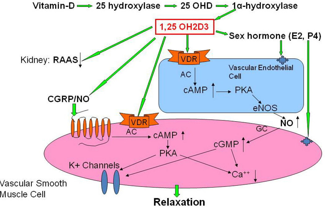
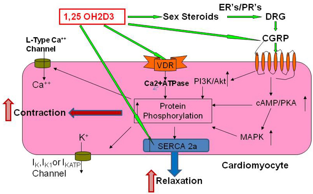
Both basic science and clinical studies support the concept that vitamin D deficiency is involved in the pathogenesis of cardiovascular and renal diseases through its association with diabetes, obesity, and hypertension. Understanding the underlying mechanisms may provide a rationale for advocating adequate intake of vitamin D and calcium in all populations, thereby preventing many chronic diseases. This review explores the effect of vitamin D deficiency in the development of cardiovascular and renal diseases, and the role of vitamin D supplementation on cardiovascular outcomes. In addition, it highlights the importance of vitamin D intake for the prevention of adverse long-term health consequences, and in ways to facilitate the management of cardiovascular disease. This is particularly true for African American and postmenopausal women, who are at added risk for cardiovascular disease. We suggest that the negative cardiovascular effects of low vitamin D in postmenopausal women could be improved by a combined treatment of vitamin D and sex steroids acting through endothelium-dependent and/or -independent mechanisms, resulting in the generation of nitric oxide and calcitonin gene-related peptide (CGRP).


Figures

Figure 1
Figure 1
Proposed schematic diagram for involvement of vitamin D in the regulation of vascular functions. Vitamin D regulates estrogen (E2) synthesis in gonads by maintaining calcium homeostasis, and E2 binds to its receptors on endothelial cells and smooth muscle cells to regulate vascular functions. In addition, vitamin D activates vitamin D receptors (VDR) on endothelial cells and vascular smooth muscle cells to regulate endogenous vasodilators, including prostaglandins (PGs), nitric oxide (NO) and calcitonin gene related peptide (CGRP). Vitamin D deficiency results in NO-mediated endothelial dysfunction, and supplementation of vitamin D attenuates this symptom. In addition, vitamin D plays a pivotal role in the down-regulation of renin-angiotensin-aldosterone system (RAAS) by the kidney. Lower vitamin D levels may lead to an impaired RAAS and abnormalities in vascular relaxation that eventually lead to the development of hypertension.
Figure 2
Figure 2
Proposed schematic diagram for involvement of vitamin D in the myocardial contractility. Cardiomyocytes express vitamin D receptors (VDR), estrogen receptors (ER’s), progesterone receptors (PR’s) and CGRP receptors. Vitamin D regulates myocyte function through the biosynthesis of steroid hormone and CGRP. In addition, vitamin D has both genomic and nongenomic effects on the cardiomyocyte functions. VDR has been found to localize to t-tubules in the heart, and is in association with Serca-2, the sarcoplasmic reticulum Ca2+-ATPase, suggesting a role of VDR in regulating cardiac contractility by a direct interaction with Serca-2. Thus, vitamin D exerts an immediate effect on signal transduction mediators and ion channels in the cardiomyocyte, and plays an important role in not only heart structure, but also regulation of its function.
Figure 3
Figure 3
Effect of chronic depletion of estrogens on the vascular vitamin D metabolizing enzyme, 1α-hydroxylase (1α-OHD) protein expression. Representative immunoblot and densitometric analysis data for 1α-OHD were depicted in figure 3. Groups of wild type control (WT) and follicle stimulating hormone receptor knockout female mice (FORKO; a model of hypertension and menopause,) were sacrificed and thoracic aortas were used for the westernblot analysis. The values are means ± SE for 3 animals in each group. P<0.05 compared to WT group.
Figure 4
Figure 4
Effect of chronic depletion of estrogens on the vascular vitamin D receptor (VDR) protein expression. Representative immunoblot and densitometric analysis data for VDR were depicted in figure 4. Groups of wild type control (WT) and follicle stimulating hormone receptor knockout female mice (FORKO; a model of hypertension and menopause, ) were sacrificed and thoracic aortas were used for the westernblot analysis. The values are means ± SE for 3 animals in each group. P<0.05 compared to WT group.
Figure 5
Figure 5
Effect of chronic depletion of estrogens on vascular neuronal nitric oxide synthase enzyme (nNOS) protein expression. Representative immunoblot and densitometric analysis data for vascular nNOS alpha were depicted in figure 5. Groups of wild type control (WT) and follicle stimulating hormone receptor knockout female mice (FORKO; a model of hypertension and menopause, ) were sacrificed and thoracic aortas were used for the westernblot analysis. The values are means ± SE for 3 animals in each group. P<0.05 compared to WT group.
Figure 6
Figure 6
Effect of chronic depletion of estrogens on vascular CGRP receptor component, calcitonin like receptor (CRLR) protein expression. Representative immunoblot and densitometric analysis data for vascular CRLR were depicted in figure 6. Groups of wild type control (WT) and follicle stimulating hormone receptor knockout female mice (FORKO; a model of hypertension and menopause, ) were sacrificed and thoracic aortas were used for the westernblot analysis. The values are means ± SE for 3 animals in each group. P<0.05 compared to WT group.
Figure 7
Figure 7
Effect of chronic depletion of estrogens on vascular CGRP receptor component, receptor activity modifying protein 1 (RAMP1) protein expression. Representative immunoblot and densitometric analysis data for vascular RAMP1 were depicted in figure 7. Groups of wild type control (WT) and follicle stimulating hormone receptor knockout female mice (FORKO; a model of hypertension and menopause, ) were sacrificed and thoracic aortas were used for the westernblot analysis. The values are means ± SE for 3 animals in each group. P<0.05 compared to WT group.

References

    1. Wilson PW, Kannel WB, Sibershatz H, D’Agostino RB. Clustering of metabolic factors and coronary heart disease. Arch Intern Med. 1999;159:1104–1109. - PubMed
    1. Kannel WB. Blood pressure as a cardiovascular risk factor: prevention and treatment. J Am Med Assoc. 1996;275:1571–1576. - PubMed
    1. Hajjar I, Kotchen TA. Trends in prevalence, awareness, treatment, and control of hypertension in the United States, 1988–2000. JAMA. 2003;290:199–206. - PubMed
    1. Androne AS, Hryniewicz K, Hudaihed A. Comparison of metabolic vasodilation in response to exercise and ischemia and endothelium-dependent flow-mediated dilation in African-American versus non-African-American patients with chronic heart failure. Am J Cardiol. 2006;97:685–689. - PubMed
    1. Rees M, Stevenson J. British Menopause Society Council. Primary prevention of coronary heart disease in women. Menopause Int. 2008;14:40–45. - PubMed

Publication types

MeSH terms