Case Reports
. 2013 Jan;141(1):61-72.
doi: 10.1085/jgp.201210899.

Induced pluripotent stem cells used to reveal drug actions in a long QT syndrome family with complex genetics

Affiliations
Case Reports

Induced pluripotent stem cells used to reveal drug actions in a long QT syndrome family with complex genetics

Cecile Terrenoire et al. J Gen Physiol. 2013 Jan.

Abstract

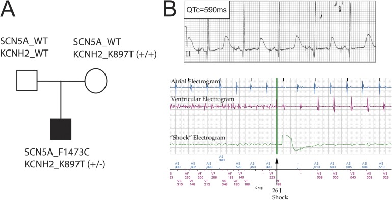
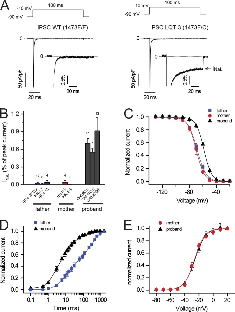
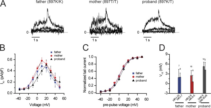
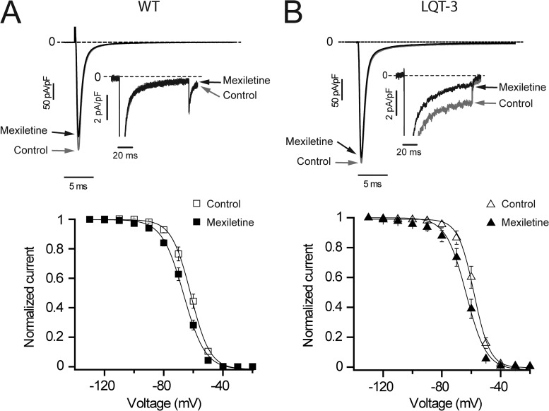
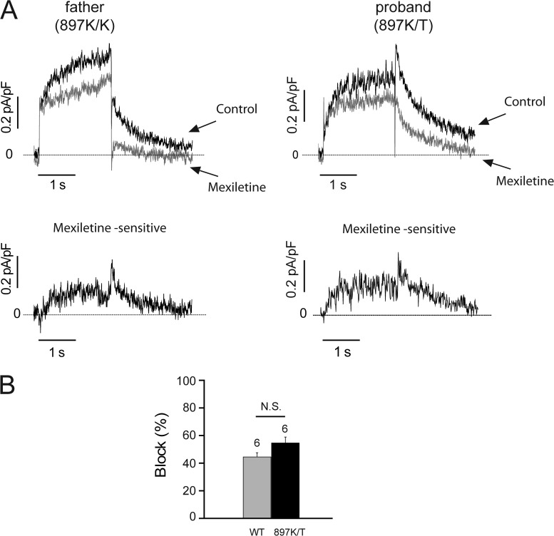
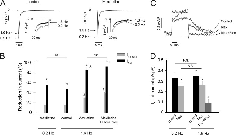
Understanding the basis for differential responses to drug therapies remains a challenge despite advances in genetics and genomics. Induced pluripotent stem cells (iPSCs) offer an unprecedented opportunity to investigate the pharmacology of disease processes in therapeutically and genetically relevant primary cell types in vitro and to interweave clinical and basic molecular data. We report here the derivation of iPSCs from a long QT syndrome patient with complex genetics. The proband was found to have a de novo SCN5A LQT-3 mutation (F1473C) and a polymorphism (K897T) in KCNH2, the gene for LQT-2. Analysis of the biophysics and molecular pharmacology of ion channels expressed in cardiomyocytes (CMs) differentiated from these iPSCs (iPSC-CMs) demonstrates a primary LQT-3 (Na(+) channel) defect responsible for the arrhythmias not influenced by the KCNH2 polymorphism. The F1473C mutation occurs in the channel inactivation gate and enhances late Na(+) channel current (I(NaL)) that is carried by channels that fail to inactivate completely and conduct increased inward current during prolonged depolarization, resulting in delayed repolarization, a prolonged QT interval, and increased risk of fatal arrhythmia. We find a very pronounced rate dependence of I(NaL) such that increasing the pacing rate markedly reduces I(NaL) and, in addition, increases its inhibition by the Na(+) channel blocker mexiletine. These rate-dependent properties and drug interactions, unique to the proband's iPSC-CMs, correlate with improved management of arrhythmias in the patient and provide support for this approach in developing patient-specific clinical regimens.


Figures

Figure 1.
Figure 1.
Clinical and genetic profile of the LQTS family. (A) Genotype of SCN5A and KCNH2 of each family member. SCN5A_1473F/F is referred to as WT SCN5A in the text. See text (Results section hERG channel activity in iPSC-CMs) for definitions of KCNH2 variants. (B, top) Surface electrocardiography lead II recorded from the LQT-3 proband before this study. QTc, QT interval corrected for heart rate. (bottom) Example of ICD detection and termination of ventricular tachyarrhythmia in proband.
Figure 2.
Figure 2.
Characterization of iPSC clones from each family member. (A) Karyotype in one clone from each family individual (father, HR-I-15; mother, HR-II-9; proband, OA6 17Cr8). (B) Expression of pluripotency markers in one clone from each family individual (father, HR-I-15; mother, HR-II-9; proband, OA6 17Cr8). (C) Teratoma assay indicated trilineage in vivo differentiation of the proband iPSC clone (OA6 17Cr8). Bars, 200 μm.
Figure 3.
Figure 3.
Action potential phenotypes in iPSC-CMs. (A) In a total of 22 cells, 2 different types of action potentials were recorded: atrial-like (left; mean of two cells) and ventricular-like (right; mean of 20 cells). Action potentials were elicited with 3-ms suprathreshold stimuli at 0.2 Hz. (B) Distribution of CMs according to their action potential phenotype: out of 22 cells, 2 cells were atrial-like and 20 cells were ventricular-like.
Figure 4.
Figure 4.
Na+ channel currents in response to adult ventricular action potential waveforms. (A and B) Adult ventricular action potential waveforms were computed (ten Tusscher et al., 2004) and applied as command waveforms in whole-cell voltage-clamp mode on iPSC-CMs from father (A) and proband (B) using recording solutions to reveal Na+ channel currents. Mexiletine-dissected Na+ channel currents are shown, averaged for four (A) and five (B) cells.
Figure 5.
Figure 5.
Biophysical properties of Na+ channels in iPSC-CMs. (A) TTX (50 µM)-sensitive averaged Na+ current traces recorded from father (WT, n = 27, three clones; left) and proband (LQT-3, n = 41, clone OA6 9Cr8; right) iPSC-CMs at −10 mV (100-ms pulses at 0.2 Hz from a −90-mV holding potential). Dashed lines, zero current. (insets) INa normalized to peak current reveal the presence of INa late current (INaL; arrow) in LQT-3 but not in WT iPSC-CMs. (B) Percentage of INaL with respect to peak current measured in three clones from the father, two clones from the mother, and three clones from the proband. (All three LQT-3 clones are P < 0.01 vs. each WT clone). The number of cells tested is indicated above each bar. (C) Steady-state availability in LQT-3 (proband, n = 27, averaged from two clones) and WT iPSC-CMs (father, n = 23, averaged from three clones; mother, n = 3, one clone; both are P < 0.001 vs. LQT-3). For each individual, data obtained from each clone are detailed in Table 1. (D) Recovery from inactivation in LQT-3 (proband, n = 6, clone OA6 9Cr8) and WT iPSC-CMs (father, n = 4, clone HR-I-2R 2Cr). Half-time in WT iPSC-CMs is P < 0.01 versus LQT-3. (E) Activation curve in LQT-3 (proband, n = 3, clone OA6 9Cr8) and WT iPSC-CMs (mother, n = 3, clone HR-II-9). V1/2 in WT iPSC-CMs is not significantly different from LQT-3. Data are shown as means ± SEM.
Figure 6.
Figure 6.
hERG K897T polymorphism does not alter biophysical properties of hERG channels in iPSC-CMs. (A) IKr currents recorded from father (897K/K), mother (897T/T), and proband (897K/T) iPSC-CMs during 2-s pulses (0.1 Hz) to −40, −10, and 20 mV and for 2 s after return to a −40-mV holding potential. Dashed lines, zero current. (B and C) Current versus test pulse voltage. (B) Current measured at 2 s during test pulse (n = 4–15). (C) Normalized tail current measured at −40 mV (n = 12–22). (D) Bar graph summary showing IKr midpoint of activation (V1/2) for each clone tested and calculated from a Boltzmann fit of normalized tail current. Data are shown as means ± SEM.
Figure 7.
Figure 7.
Mexiletine corrects F1473C-altered Na+ channel inactivation. (A and B, top) Averaged INa current traces recorded at −10 mV (100-ms pulses applied at 0.2 Hz from a −90-mV holding potential) in father (A; WT, n = 5, clone HR-I-2R 2Cr) and proband (B; LQT-3, n = 18, clone OA6 9Cr8) iPSC-CMs in control conditions (gray traces and arrows) and in the presence of 50 µM mexiletine (black traces and arrows) at low and high (insets) gain. (insets) INa at high gain shows the absence of mexiletine-sensitive INaL in father’s iPSC-CMs (A) but reveals the presence of INaL in LQT-3 iPSC-CMs, which was significantly blocked by 50 µM mexiletine (B, arrow). (A and B, bottom) Steady-state availability in the absence (open symbols) and presence (closed symbols) of 50 µM mexiletine in WT (A; n = 5, clone HR-I-2R 2Cr) and LQT-3 (B; n = 8, clone OA6 9Cr8) cells. Data are shown as means ± SEM.
Figure 8.
Figure 8.
Mexiletine blocks IKr channels. (A, top) IKr current traces recorded in father and proband iPSC-CMs during a 2-s depolarizing test pulse to 30 mV followed by a 2-s repolarizing pulse to −40 mV (holding potential was −40 mV) at 0.1 Hz in control conditions (black traces and arrows) and in the presence of 50 µM mexiletine (gray traces and arrows). 30 µM chromanol 293B was added to block IKs. (bottom) Mexiletine (50 µM)-sensitive potassium currents in iPSC-CMs subtracted from the top recordings. Dashed lines, zero current. (B) 50 µM mexiletine significantly blocked IKr tail current at −40 mV in iPSC-CMs from the father (WT = 897K/K, P < 0.05 vs. control) and proband (897K/T, P < 0.05 vs. control). Data are shown as means ± SEM.
Figure 9.
Figure 9.
Impact of stimulation frequency on INaL and IKr in the absence and presence of drugs. (A) Averaged sodium current traces recorded at −10 mV (100-ms pulses applied from a −90-mV holding potential) in LQT-3 iPSC-CMs in control conditions (left; n = 5) and in the presence of 50 µM mexiletine (right; n = 5) at 0.2 Hz (gray traces and arrows) and at 1.6 Hz (black traces and arrows) at low and high (insets) gain. Dashed lines, zero current. (B) Percent block of INa peak current (INa peak) and INa late current (INaL) at 0.2 and 1.6 Hz by 50 µM mexiletine alone (n = 6) and 50 µM mexiletine plus 5 µM flecainide (n = 3). One-way ANOVA followed by Tukey’s test: *, P < 0.01 versus paired INa peak; #, P < 0.01 versus INa peak at 0.2 Hz in the presence of mexiletine; δ, P < 0.01 versus INaL at 0.2 Hz in the presence of mexiletine. (C) Block of IKr channels by 50 µM mexiletine (Mex) and 5 µM flecainide (Flec) at 1.6 Hz in WT iPSC-CMs depolarized to 30 mV for 200 ms, from a holding potential of −40 mV, and repolarized at −40 mV for 200 ms. (D) Change in IKr tail current density measured at −40 mV after 200-ms test pulses at 30 mV in control condition (at 0.2 and 1.6 Hz), in the presence of 50 µM mexiletine and after the addition of 5 µM flecainide. *, P < 0.05 versus paired control; #, P < 0.05 versus mexiletine at 1.6 Hz. Data are shown as means ± SEM.

Comment in

References

    1. An R.H., Bangalore R., Rosero S.Z., Kass R.S. 1996. Lidocaine block of LQT-3 mutant human Na+ channels. Circ. Res. 79:103–108 10.1161/01.RES.79.1.103 - DOI - PubMed
    1. Anson B.D., Ackerman M.J., Tester D.J., Will M.L., Delisle B.P., Anderson C.L., January C.T. 2004. Molecular and functional characterization of common polymorphisms in HERG (KCNH2) potassium channels. Am. J. Physiol. Heart Circ. Physiol. 286:H2434–H2441 10.1152/ajpheart.00891.2003 - DOI - PubMed
    1. Bankston J.R., Sampson K.J., Kateriya S., Glaaser I.W., Malito D.L., Chung W.K., Kass R.S. 2007a. A novel LQT-3 mutation disrupts an inactivation gate complex with distinct rate-dependent phenotypic consequences. Channels (Austin). 1:273–280 - PubMed
    1. Bankston J.R., Yue M., Chung W., Spyres M., Pass R.H., Silver E., Sampson K.J., Kass R.S. 2007b. A novel and lethal de novo LQT-3 mutation in a newborn with distinct molecular pharmacology and therapeutic response. PLoS ONE. 2:e1258 10.1371/journal.pone.0001258 - DOI - PMC - PubMed
    1. Bezzina C.R., Verkerk A.O., Busjahn A., Jeron A., Erdmann J., Koopmann T.T., Bhuiyan Z.A., Wilders R., Mannens M.M., Tan H.L., et al. 2003. A common polymorphism in KCNH2 (HERG) hastens cardiac repolarization. Cardiovasc. Res. 59:27–36 10.1016/S0008-6363(03)00342-0 - DOI - PubMed

Publication types

MeSH terms