Review
. 2012 Nov 1;125(Pt 21):4951-61.
doi: 10.1242/jcs.038083.

Centrosome positioning in vertebrate development

Affiliations
Review

Centrosome positioning in vertebrate development

Nan Tang et al. J Cell Sci. .

Abstract

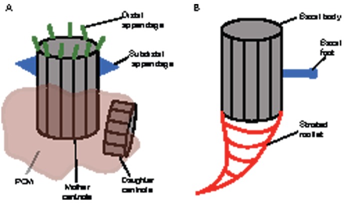
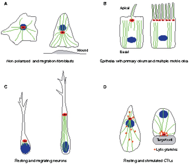
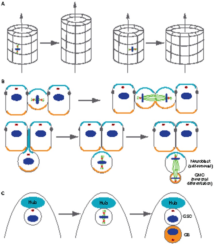
The centrosome, a major organizer of microtubules, has important functions in regulating cell shape, polarity, cilia formation and intracellular transport as well as the position of cellular structures, including the mitotic spindle. By means of these activities, centrosomes have important roles during animal development by regulating polarized cell behaviors, such as cell migration or neurite outgrowth, as well as mitotic spindle orientation. In recent years, the pace of discovery regarding the structure and composition of centrosomes has continuously accelerated. At the same time, functional studies have revealed the importance of centrosomes in controlling both morphogenesis and cell fate decision during tissue and organ development. Here, we review examples of centrosome and centriole positioning with a particular emphasis on vertebrate developmental systems, and discuss the roles of centrosome positioning, the cues that determine positioning and the mechanisms by which centrosomes respond to these cues. The studies reviewed here suggest that centrosome functions extend to the development of tissues and organs in vertebrates.


Figures

Fig. 1.
Fig. 1.
The anatomy of vertebrate centrosomes and centrioles. (A) Schematic diagram of typical centrosome and centriole organization in most vertebrate cells. Centrosomes are composed of two orthogonally oriented centrioles that are surrounded by PCM. The mother centriole is distinguished by two sets of appendages, the subdistal and distal appendages (Paintrand et al., 1992), which are thought to be required for anchoring microtubules at the centriole or forming transitional fibers that contact the cell cortex, respectively (Dawe et al., 2007). (B) Organization of the basal body (centriole) in a cell from a multiciliated epithelium. The proximal side of the basal body is associated with a matrix, which extends into specific and striated structures called rootlets (Klotz et al., 1986). PCM, pericentriolar material.
Fig. 2.
Fig. 2.
Examples of centrosome positioning that depend on cell type and cell state. (A) Fibroblasts: in non-polarized fibroblasts (left), the centrosome is located near the center of the cell and is physically linked to the nucleus, with microtubules radiating from the centrosome to the cell cortex. During wound healing (right), the fibroblast centrosome often becomes oriented between the nucleus and the leading edge. (B) Epithelial cells: in polarized epithelial cells (left), centrosomes are located on the apical surface of the cell. The apical localization of the centrosome is accompanied by a loss of radial microtubule organization and the formation of a predominantly apical–basal array of microtubules. The mother centriole of the centrosome becomes a basal body, which gives rise to a primary cilium. In multiciliated epithelial cells (right), hundreds of centrioles are assembled at once in a single cell, leading to the formation of multiple cilia. (C) Neurons: in resting neurons (left), the centrosome is found in close proximity to the neurite that becomes the axon. In migrating neurons (right), the centrosome is positioned ahead of the nucleus, with microtubules connecting the centrosome and the nucleus. (D) Lymphocytes: in cytotoxic T lymphocytes (CTLs) that are not interacting with a target (left), the centrosome is located near the nucleus, and lytic granules are distributed all along the microtubules. After the CTL is stimulated (right), the centrosome directs the delivery of lytic granules by moving along microtubules to the plasma membrane and then to the point of secretion for releasing lytic granules to the immunological synapse. Red ovals indicate the centrosome, with centrioles indicated as black lines within. Blue ovals indicate nuclei and green lines indicate microtubules.
Fig. 3.
Fig. 3.
Spindle orientation mediated by centrosome–cortex interactions has functional roles and effects on tubular morphogenesis (A) and specification of cell fate (B and C). (A) Mitotic spindle orientation with respect to the longitudinal axis of an epithelial tube has a major effect on its shape. When the spindle orientation is parallel to the longitudinal axis of the tube (shown on the left), this cell division only increases tube length but not the circumference. By contrast, when the spindle orientation is perpendicular to the longitudinal axis of the tube (shown on the right), the cell division only increases tube circumference but not the length. (B) Intrinsic asymmetrically localized cortical cues (represented by the orange and turquoise coloring) determine the different fates of daughter cells in neuroblasts. Symmetrical divisions (top row), in which the mitotic spindles are perpendicular to the apical–basal axis, generate two identical daughter cells that inherit both apical and basal cortical cues. Asymmetric cell divisions (bottom row), in which mitotic spindles are parallel to the apical–basal axis, generate two daughter cells that inherit either apical or basal cortical cues. One daughter cell self-renews to maintain the pool of neuroblasts, whereas the other differentiates to populate the central nervous system. (C) Extrinsic polarity cues determine the different fates of daughter cells in the Drosophila male germline stem cell (GSC). During cell division, the mitotic spindle forms perpendicular to the interface with the stem cell niche (Hub), such that one daughter cell retains contact with the niche and its sustaining signals, whereas the other daughter cell loses contact with the niche and initiates differentiation (gonialblast, shown in orange). GMC, ganglion mother cell; GB, gonialblast.

Similar articles

Cited by

References

    1. Abal M., Keryer G., Bornens M. (2005). Centrioles resist forces applied on centrosomes during G2/M transition. Biol. Cell 97, 425–434 10.1042/BC20040112 - DOI - PubMed
    1. Albrecht–Buehler G. (1977). Phagokinetic tracks of 3T3 cells: parallels between the orientation of track segments and of cellular structures which contain actin or tubulin. Cell 12, 333–339 10.1016/0092-8674(77)90109-X - DOI - PubMed
    1. Azimzadeh J., Bornens M. (2007). Structure and duplication of the centrosome. J. Cell Sci. 120, 2139–2142 10.1242/jcs.005231 - DOI - PubMed
    1. Azimzadeh J., Wong M. L., Downhour D. M., Sánchez Alvarado A., Marshall W. F. (2012). Centrosome loss in the evolution of planarians. Science 335, 461–463 10.1126/science.1214457 - DOI - PMC - PubMed
    1. Baena–López L. A., Baonza A., García–Bellido A. (2005). The orientation of cell divisions determines the shape of Drosophila organs. Curr. Biol. 15, 1640–1644 10.1016/j.cub.2005.07.062 - DOI - PubMed

Publication types

Substances