. 2013 Jan 15;110(3):1124-9.
doi: 10.1073/pnas.1212113110. Epub 2012 Dec 31.

Robust and tunable circadian rhythms from differentially sensitive catalytic domains

Affiliations

Robust and tunable circadian rhythms from differentially sensitive catalytic domains

Connie Phong et al. Proc Natl Acad Sci U S A. .

Abstract

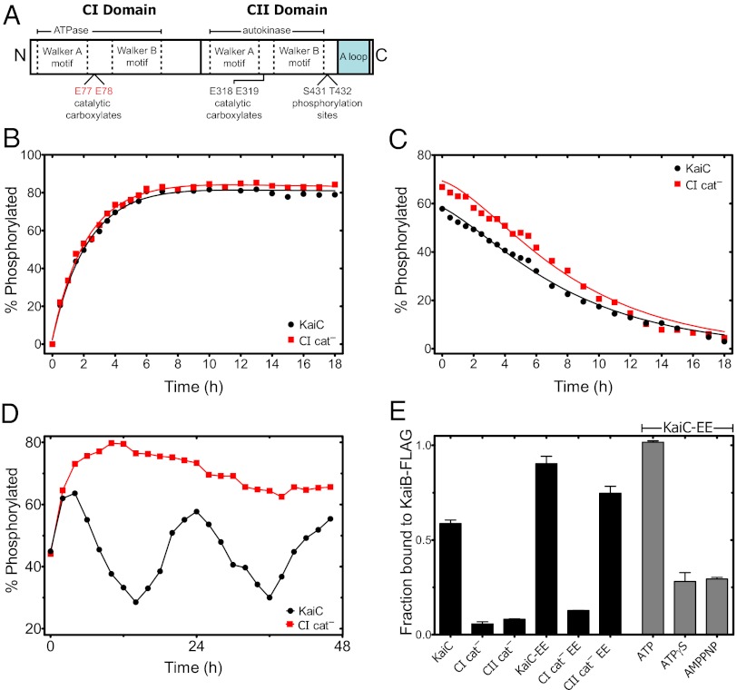
Circadian clocks are ubiquitous biological oscillators that coordinate an organism's behavior with the daily cycling of the external environment. To ensure synchronization with the environment, the period of the clock must be maintained near 24 h even as amplitude and phase are altered by input signaling. We show that, in a reconstituted circadian system from cyanobacteria, these conflicting requirements are satisfied by distinct functions for two domains of the central clock protein KaiC: the C-terminal autokinase domain integrates input signals through the ATP/ADP ratio, and the slow N-terminal ATPase acts as an input-independent timer. We find that phosphorylation in the C-terminal domain followed by an ATPase cycle in the N-terminal domain is required to form the inhibitory KaiB•KaiC complexes that drive the dynamics of the clock. We present a mathematical model in which this ATPase-mediated delay in negative feedback gives rise to a compensatory mechanism that allows a tunable phase and amplitude while ensuring a robust circadian period.


Conflict of interest statement

The authors declare no conflict of interest.

Figures

Fig. 1.
Fig. 1.
The period of the circadian oscillator is robust to varying input signals. (A) Schematic of the KaiABC oscillator. A purified posttranslational protein circuit generates circadian rhythms in KaiC phosphorylation in the presence of ATP. Active KaiA promotes autophosphorylation on initially unphosphorylated KaiC (U-KaiC) first on Thr432 and then, on Ser431. Ser431-phosphorylated KaiC (S-KaiC) forms KaiB•KaiC complexes that inhibit KaiA and promote system-wide dephosphorylation, driving stable oscillations. The phase of the oscillation can be shifted by metabolic signaling through the ATP/ADP ratio. (B) KaiC autophosphorylation in KaiA-KaiC reactions at various ATP/ADP conditions (colored symbols); all buffers have 10 mM total nucleotide (similar data previously published in ref. 6). (C) KaiABC in vitro reactions in buffers at various ATP/ADP conditions; 10 mM total nucleotide. Oscillations with a robust period persist under the conditions tested. (D) Period of the oscillations shown in C determined by fitting the data after the first trough to a sinusoidal function. Error bars indicate the SE of the least squares fit. Dashed lines indicate linear regressions of the data to show trend. (E) Peak and trough heights of the oscillations shown in C determined by averaging the peaks and troughs after the first trough. Error bars indicate SDs. Dashed lines indicate linear regressions of the data to show trend.
Fig. 2.
Fig. 2.
CI ATPase activity is required for KaiB•KaiC complex formation. (A) KaiC is organized into tandem catalytic domains, CI and CII, both containing conserved Walker A and Walker B motifs that flank pairs of catalytic residues. CII has autokinase activity, which is promoted by interactions between the C-terminal A-loop region and KaiA. In the CI cat mutant protein, the catalytic carboxylates Glu77 and Glu78 are both mutated to glutamine. (B) Autophosphorylation of KaiC WT (black circles) and CI cat mutant (red squares) in the presence of KaiA. (C) Autodephosphorylation of KaiC WT and CI cat mutant. Highly phosphorylated protein was prepared by incubation with KaiA and then subsequently removed by immunoprecipitation to initiate dephosphorylation. Solid lines in B and C are fits to a four-state kinetic model of phosphoform interconversion. (D) Phosphorylation dynamics of KaiC WT and CI cat mutant protein in the presence of KaiA and KaiB. (E) Levels of WT KaiC, CI cat, and a CII domain catalytic mutant (CII cat, E318Q) protein coimmunoprecipitating with KaiB-FLAG in the presence of KaiA after incubation for 10 h. Data are also shown for phosphomimetic (S431E; T432E) KaiC in various backgrounds of catalytic site mutations: WT (KaiC-EE), CI cat (CI EE), and CII cat (CII EE; black bars). KaiC-EE coimmunoprecipitating with KaiB-FLAG after incubation for 8–10 h in buffers with the nonhydrolyzable ATP analogs ATPγS and adenylyl imidodiphosphate (AMPPNP) (gray bars). Error bars indicate SEs from three replicates.
Fig. 3.
Fig. 3.
CI ATPase and CII autokinase activities are differentially regulated by ADP. (A) KaiA-stimulated CII domain autophosphorylation in the CI cat mutant protein at various ATP/ADP conditions (colored symbols); all buffers have 1 mM ATP. (B) Kinase rate constants with error bars indicating SEs of fits (△, kCII kinase) obtained from data in A. CI ATPase rate constants (●, kCI ATPase) measured as total ADP production by the CI fragment (KaiC residues 1–247) in buffers at various ATP/ADP levels; all buffers have 1 mM ATP. Error bars indicate SEMs of at least three measurements. Dashed lines indicate fits to a competitive inhibition model, k{[ATP]/([ATP] + KI[ADP])}: KI/CII kinase = 0.96 ± 0.18 and KI/CI ATPase = 0.10 ± 0.03. (C) Amount of phosphomimetic KaiC-EE coimmunoprecipitating with KaiB-FLAG as a function of time under various total protein concentrations with equimolar amounts of KaiB-FLAG and KaiC-EE (0.5× = 1.75 μM, 1× = 3.5 µM, 2× = 7 μM, and 4× = 14 μM). Dashed lines indicate predicted assembly curves for KaiB•KaiC complex assembly rates set by bimolecular collisions for each concentration. (D) Amount of phosphomimetic KaiC-EE coimmunoprecipitating with KaiB-FLAG as a function of time under various ATP/ADP conditions. Solid lines in C and D indicate fits to a pseudofirst-order reversible binding reaction (SI Text). (E) Schematic of requirements for KaiB•KaiC complex assembly. U-KaiC first autophosphorylates in the CII domain on Thr432 and then Ser431. Autokinase activity in CII depends on KaiA and the ATP/ADP input signal. After Ser431 phosphorylation is achieved, KaiC undergoes a catalytic cycle in the CI domain, causing a hypothetical structural rearrangement to occur and permitting a stable KaiB•KaiC complex to form. ATPase activity in CI is insensitive to the ATP/ADP input signal.
Fig. 4.
Fig. 4.
Mathematical model integrating roles of CI and CII predicts a robust period. (A) Model A consists of a modified reaction network including a slow binding step between Ser431-phosphorylated KaiC and KaiB that is invariant to ATP/ADP (red arrow) in addition to kinase reactions sensitive to ATP/ADP (black arrows) and phosphatase reactions (gray arrows). Numerical integration of this model reproduces oscillations of KaiC phosphorylation with a robust period over a range of ATP/ADP conditions. (B) Model B neglects the role of CI and assumes that KaiB binding occurs much faster than changes in CII phosphorylation. Numerical integration of this model shows that the period elongates and oscillations lose stability at low ATP/ADP conditions. (C) Period of the oscillations at various ATP/ADP for models A (red symbols) and B (blue symbols) determined by fitting the simulated trajectory to a sinusoidal function. Periods under conditions where oscillations are unstable were estimated by fitting to initial transient oscillations and are represented by dashed lines. All periods have been scaled relative to an assumed 24-h period for the 100% ATP condition for each model. (D) Phase response curve showing relationship between time of 50% ADP pulse and the resulting phase advance or delay (model A, red symbols) compared with the phase response from model B (blue symbols) and from in vitro (orange triangles) and in vivo (green squares) experimental data. Comparisons were made by scaling the period of the models to 24 h and shifting the data so that peak phosphorylation occurs at CT 12.

Similar articles

Cited by

References

    1. Winfree AT. The Geometry of Biological Time. 2nd Ed. New York: Springer; 2000.
    1. Ouyang Y, Andersson CR, Kondo T, Golden SS, Johnson CH. Resonating circadian clocks enhance fitness in cyanobacteria. Proc Natl Acad Sci USA. 1998;95(15):8660–8664. - PMC - PubMed
    1. Scheer FA, Hilton MF, Mantzoros CS, Shea SA. Adverse metabolic and cardiovascular consequences of circadian misalignment. Proc Natl Acad Sci USA. 2009;106(11):4453–4458. - PMC - PubMed
    1. Martino TA, et al. Circadian rhythm disorganization produces profound cardiovascular and renal disease in hamsters. Am J Physiol Regul Integr Comp Physiol. 2008;294(5):R1675–R1683. - PubMed
    1. Nakajima M, et al. Reconstitution of circadian oscillation of cyanobacterial KaiC phosphorylation in vitro. Science. 2005;308(5720):414–415. - PubMed

Publication types

MeSH terms

Substances

LinkOut - more resources