. 2013 Jan 15;110(3):936-41.
doi: 10.1073/pnas.1212070110. Epub 2012 Dec 31.

DnaA binding locus datA promotes DnaA-ATP hydrolysis to enable cell cycle-coordinated replication initiation

Affiliations

DnaA binding locus datA promotes DnaA-ATP hydrolysis to enable cell cycle-coordinated replication initiation

Kazutoshi Kasho et al. Proc Natl Acad Sci U S A. .

Abstract

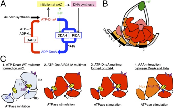
The initiation of chromosomal DNA replication is rigidly regulated to ensure that it occurs in a cell cycle-coordinated manner. To ensure this in Escherichia coli, multiple systems regulate the activity of the replication initiator ATP-DnaA. The level of ATP-DnaA increases before initiation after which it drops via DnaA-ATP hydrolysis, yielding initiation-inactive ADP-DnaA. DnaA-ATP hydrolysis is crucial to regulation of initiation and mainly occurs by a replication-coupled feedback mechanism named RIDA (regulatory inactivation of DnaA). Here, we report a second DnaA-ATP hydrolysis system that occurs at the chromosomal site datA. This locus has been annotated as a reservoir for DnaA that binds many DnaA molecules in a manner dependent upon the nucleoid-associated factor IHF (integration host factor), resulting in repression of untimely initiations; however, there is no direct evidence for the binding of many DnaA molecules at this locus. We reveal that a complex consisting of datA and IHF promotes DnaA-ATP hydrolysis in a manner dependent on specific inter-DnaA interactions. Deletion of datA or the ihf gene increased ATP-DnaA levels to the maximal attainable levels in RIDA-defective cells. Cell-cycle analysis suggested that IHF binds to datA just after replication initiation at a time when RIDA is activated. We propose a model in which cell cycle-coordinated ATP-DnaA inactivation is regulated in a concerted manner by RIDA and datA.


Conflict of interest statement

The authors declare no conflict of interest.

Figures

Fig. 1.
Fig. 1.
datA-dependent DnaA-ATP hydrolysis. (A) Schematic presentation of the chromosomal loci and structures of oriC, dnaA, datA, DARS1/2, and ter. (Left) The location of each site on the E. coli chromosome is indicated. (Right) Open bars indicate the oriC-, datA-, and DARS1-containing fragments used in this study: respectively, FK-9, datA WT, and FK7-7. Arrowheads represent DnaA binding sites that match the 9-mer consensus sequence completely (black) or contain a mismatch(es) (dotted). R5, I1–3, τ1–2, and C1–3 in oriC are low-affinity ATP-DnaA binding sites (1, 4, 9). DnaA boxes 1–5 in datA and DnaA boxes I–III in DARS1 are displayed similarly. IBS (green bars) and AT-rich repeats that facilitate duplex unwinding (AT repeats; purple bars) are also indicated. (B–D) In vitro reconstitution of datA-dependent DnaA-ATP hydrolysis. [α-32P]ATP-DnaA (1 pmol) was incubated under various conditions, and then analyzed by Thin-layer chromatography (TLC). The proportions of ADP-DnaA to total ATP/ADP-DnaA molecules are indicated as percentages (%). (B) ATP-DnaA was incubated at 30 °C for 10 min with the indicated amounts of datA WT (datA) (●, ○) or FK-9 (oriC) (▲, △) in the presence (●, ▲) or absence (○, △) of IHF (0.2 pmol). (C) ATP-DnaA was incubated with the indicated amounts of either IHF (●, ○) or HU (▲, △) in the presence (0.05 pmol) of datA WT (●, ▲) or FK-9 (○, △). (D) Reaction time course was analyzed at 0 °C (○, △) or 30 °C (●, ▲) in the presence of IHF (0.2 pmol) and either datA WT (0.05 pmol) (●, ○) or FK-9 (▲, △). (E) In vitro reconstitution of the DnaA cycle using datA and DARS1. DnaA activity was assessed using an in vitro replication system. In the first stage, IHF (0.4 pmol) and ATP-DnaA or ADP-DnaA (2 pmol) were incubated at 30 °C for 10 min in buffer (25 µL) containing (red bars) or excluding (black bars) datA WT, followed by the addition of DpnII and incubation at 30 °C for 5 min to digest datA DNA. In the second stage, the indicated amounts of DARS1 DNA (FK7-7, 1 µL) were added to samples (27 µL) that had contained ATP-DnaA and datA WT (red bars) or ADP-DnaA (black bars) in the first stage, followed by further incubation at 30 °C for 15 min. After the first- and second-stage reactions, portions (5 µL) were withdrawn, and DnaA initiator activity was analyzed in vitro using a minichromosome replication system in a crude protein extract. In the second stage, subDnaAbox2 (blue bar), a DnaA box 2-substituted derivative of datA that is inactive in DDAH, was used as a negative control (see also Fig. 2B). In the second stage, DARS1 mutant ΔCore (FK7-21), which contains a deletion of DnaA boxes I–III and is inactive in DnaA-nucleotide exchange, was used as a negative control. Error bars represent the SD from two independent experiments.
Fig. 2.
Fig. 2.
DnaA and IHF binding sites for DDAH. (A) The open bar at the top depicts the datA region, as described in Fig. 1A. datA WT DNA and its truncated derivatives (black bars) were incubated with [α-32P]ATP-DnaA (1 pmol) and IHF at 30 °C for 10 min, followed by TLC. When the datA derivatives included were 0.05 pmol, IHF included was 0.4 pmol. When the datA derivatives included were 0.1 pmol, IHF included was 0.8 pmol. Results (%) are shown to the right of the bars. n.d., not determined. (B) Similar experiments were performed using the indicated mutants of datA or oriC FK-9. subDnaAbox1–5 and subIBS contain substitutions of the corresponding DnaA box and IBS, respectively (Fig. S1 and Table S3). (C) Schematic of the relationship between IBS and DnaA box 3. delC, delD, insB, and insB+ mutations are shown with the nucleotide numbers (25). For details in sequences, see Fig. S2. (D) An in vitro reconstituted DDAH system using datA derivatives containing a deletion or an insertion between IBS and DnaA box 3.
Fig. 3.
Fig. 3.
DnaA motifs are required for DDAH. (A) The [α-32P]ATP forms (1 pmol) of WT DnaA (●, ○) or DnaA R334A (▲, △) were incubated at 30 °C for 10 min with IHF (0.2 pmol) and the indicated amounts of datA WT (●, ▲) or oriC FK-9 (○,△). (B) The binding of WT DnaA (●, ○), DnaA R281A (▲, △), or DnaA R399A (◆, ◇) to datA (●,▲, ◆) or oriC (○, △, ◇) were similarly analyzed. (C) Similar experiments were performed using WT DnaA (●, ○) and the R285A mutant (▲, △). (D) The indicated amounts of ATP- or ADP-DnaA were incubated with datA del5 (0.15 pmol), IHF (6 pmol), and 150 ng λDNA (as a competitor) at 15 °C for 5 min, followed by EMSA and Gel-star staining. The gel image is shown in black-and-white inverted mode. IHF, IHF-bound datA. C1–C3, datA-IHF carrying 1–3 DnaA molecules. >5C, datA-IHF carrying more than five DnaA molecules. (E) The proportions of higher (>C5; ●, ○) and lower (C1–C3; ▲, △) complexes of ATP- (●, ▲) or ADP-DnaA (○, △) were determined using the data shown in D and are plotted as percentages (%).
Fig. 4.
Fig. 4.
Analyses for cellular ATP-DnaA level and IHF binding. (A) KW262-5 (rnhA::Tn3 ΔoriC) (WT), MK86 (Δhda), KX93 (ΔhdadatA), KX30 (ΔhdaihfA), KX31 (Δhda ΔihfB), and KX32 (Δhda ΔhupA) cells were grown at 37 °C in medium containing 32P. DnaA was immunoprecipitated, and recovered DnaA-bound nucleotides were analyzed by TLC. Error bars represent the SD from at least four independent experiments. (B and C) KYA018 (dnaC2) cells growing at 30 °C in supplemented M9 medium were transferred to 38 °C and incubated for 90 min. The cells were then transferred to 30 °C (time 0), incubated for 5 min, and further incubated at 38 °C; samples were withdrawn at the indicated times. The oriC, datA, and ylcC levels before (Input) and after (ChIP) immunoprecipitation using anti-IHF antiserum were determined using real-time quantitative PCR. The ChIP/Input for ylcC (%) was used as a background control for nonspecific IHF binding and was subtracted from the ChIP/Input for oriC and datA. The levels of oriC or datA relative to ter in the Input samples were also quantified using real-time quantitative PCR, and the relative ratios of oriC/ter and datA/ter are expressed relative to the ratio at 0 min (defined as 1). Relative ChIP/Input values for oriC and oriC/ter ratio (B) and the relative ChIP/Input values for datA and datA/ter ratio (C) are shown. Error bars represent the SD from at least three independent experiments.
Fig. 5.
Fig. 5.
A model for the molecular mechanism of DDAH. (A) A revised view of the DnaA activity cycle including DDAH. ATP-DnaA is hydrolyzed by two independent pathways, RIDA and DDAH. IHF stimulates initiation at oriC (9, 12, 13) and DDAH. Thus, IHF plays both positive and negative roles in initiation. DARS reactivates ADP-DnaA by ADP-to-ATP exchange. (B) A model of the structure of the DnaA-IHF-datA complex. ATP-DnaA multimers are formed on regions carrying DnaA boxes 2–4. Sharp DNA bending by IHF stimulates interaction between ATP-DnaA multimers. For simplicity, only DnaA domain III (red or pink polygon) and domain IV (orange square) are shown. (C) A model of DDAH-specific conformational change of DnaA oligomers. For simplicity, only a dimer of DnaA domain III is shown. Domain IIIa (large polygon, IIIa) contains Arg285 of the Arg-finger and Arg281 of Box VII, whereas domain IIIb (small polygon, IIIb) contains Arg334 of Sensor II. In ATP-DnaA-oriC complexes, Arg285 recognizes the neighboring DnaA-bound ATP molecule, and Arg281 supports tight inter-DnaA interactions. These events inhibit the interaction between ATP and Arg334 of Sensor II. In DnaA R281A mutant-oriC complexes, the inter-DnaA interaction is not as tight, resulting in a preponderance of the DnaA Arg334-ATP interaction. In ATP-DnaA-datA complexes, Arg281 modulates the structure of the complex and the inter-DnaA interaction is not as tight, which also allows the DnaA Arg334-ATP interaction. In ATP-DnaA-Hda complexes involved in RIDA, the DnaA-Hda-interaction is not tight, which allows ATP to interact with DnaA Arg334 and Hda Arg153.

Similar articles

Cited by

References

    1. Messer W. The bacterial replication initiator DnaA. DnaA and oriC, the bacterial mode to initiate DNA replication. FEMS Microbiol Rev. 2002;26(4):355–374. - PubMed
    1. Katayama T, Ozaki S, Keyamura K, Fujimitsu K. Regulation of the replication cycle: Conserved and diverse regulatory systems for DnaA and oriC. Nat Rev Microbiol. 2010;8(3):163–170. - PubMed
    1. Kaguni JM. Replication initiation at the Escherichia coli chromosomal origin. Curr Opin Chem Biol. 2011;15(5):606–613. - PMC - PubMed
    1. Leonard AC, Grimwade JE. Regulation of DnaA assembly and activity: Taking directions from the genome. Annu Rev Microbiol. 2011;65:19–35. - PMC - PubMed
    1. Erzberger JP, Pirruccello MM, Berger JM. The structure of bacterial DnaA: Implications for general mechanisms underlying DNA replication initiation. EMBO J. 2002;21(18):4763–4773. - PMC - PubMed

Publication types

MeSH terms