

Protective role of brain water channel AQP4 in murine cerebral malaria
- PMID: 23277579
- PMCID: PMC3549120
- DOI: 10.1073/pnas.1220566110
Protective role of brain water channel AQP4 in murine cerebral malaria
Abstract
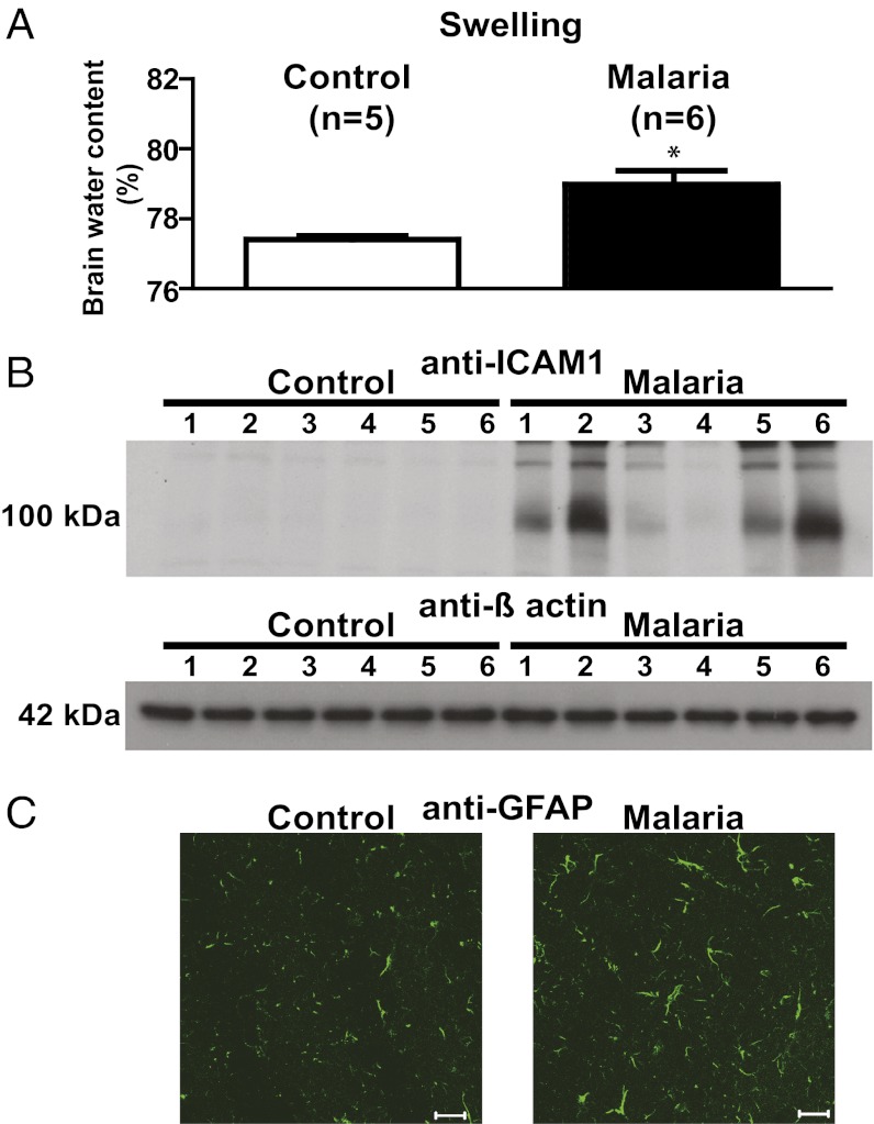
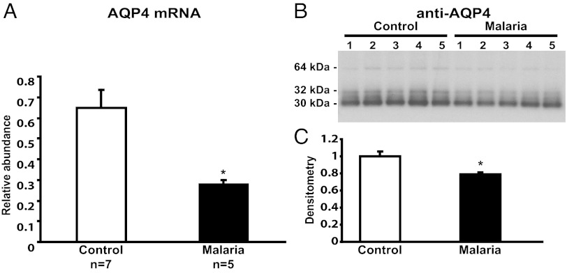
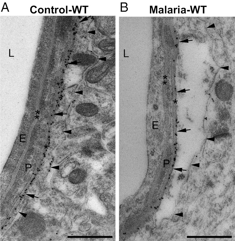
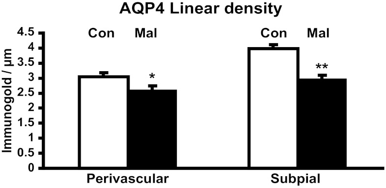
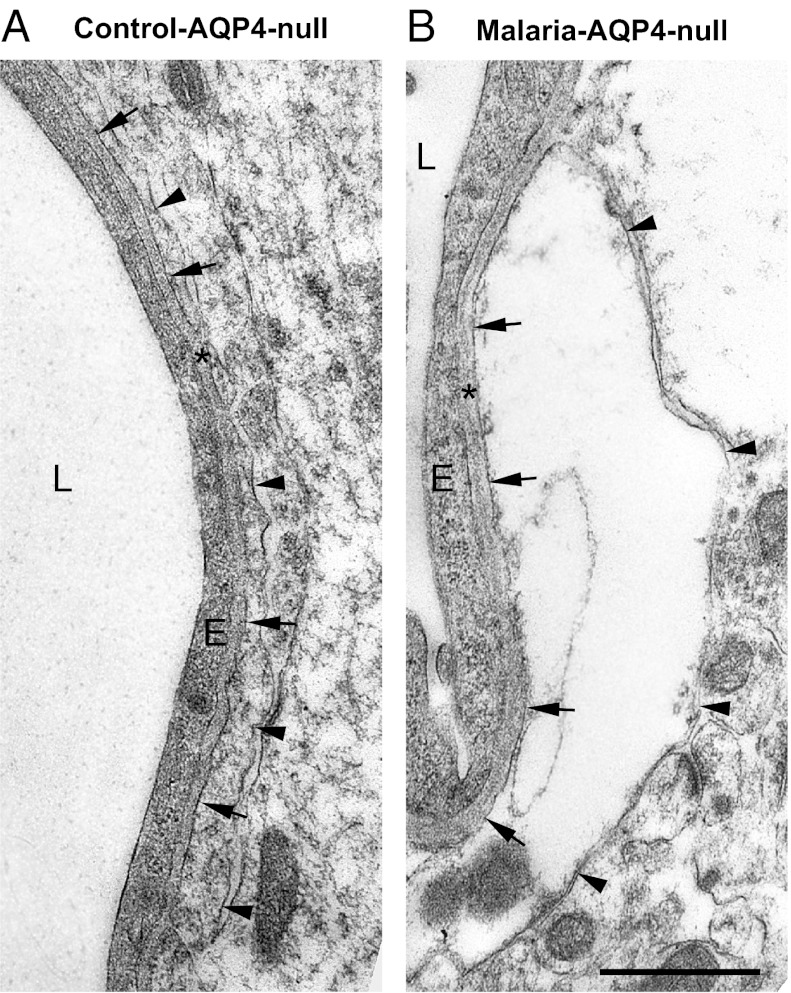
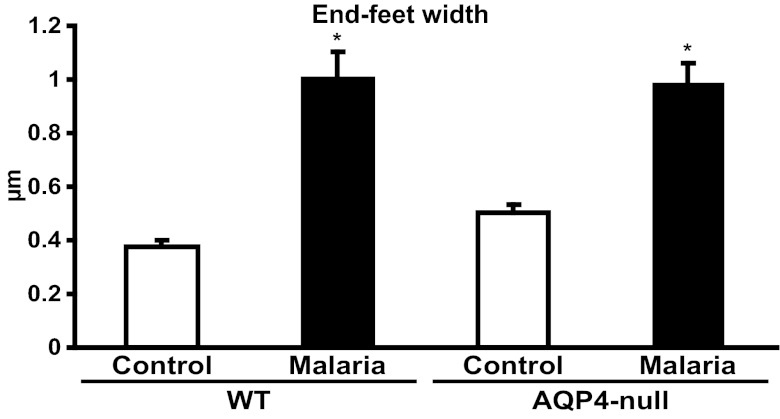
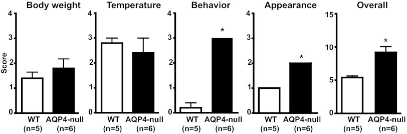
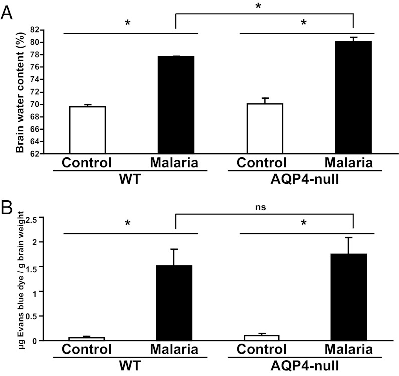
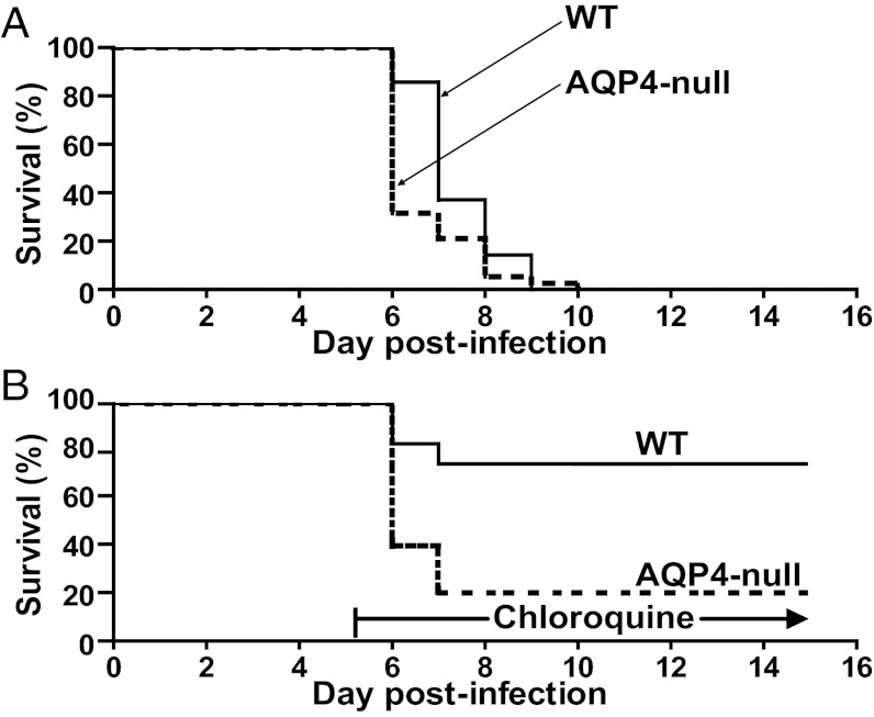
Tragically common among children in sub-Saharan Africa, cerebral malaria is characterized by rapid progression to coma and death. In this study, we used a model of cerebral malaria appearing in C57BL/6 WT mice after infection with the rodent malaria parasite Plasmodium berghei ANKA. Expression and cellular localization of the brain water channel aquaporin-4 (AQP4) was investigated during the neurological syndrome. Semiquantitative real-time PCR comparing uninfected and infected mice showed a reduction of brain AQP4 transcript in cerebral malaria, and immunoblots revealed reduction of brain AQP4 protein. Reduction of brain AQP4 protein was confirmed in cerebral malaria by quantitative immunogold EM; however, polarized distribution of AQP4 at the perivascular and subpial astrocyte membranes was not altered. To further examine the role of AQP4 in cerebral malaria, WT mice and littermates genetically deficient in AQP4 were infected with P. berghei. Upon development of cerebral malaria, WT and AQP4-null mice exhibited similar increases in width of perivascular astroglial end-feet in brain. Nevertheless, the AQP4-null mice exhibited more severe signs of cerebral malaria with greater brain edema, although disruption of the blood-brain barrier was similar in both groups. In longitudinal studies, cerebral malaria appeared nearly 1 d earlier in the AQP4-null mice, and reduced survival was noted when chloroquine rescue was attempted. We conclude that the water channel AQP4 confers partial protection against cerebral malaria.
Conflict of interest statement
The authors declare no conflict of interest.
Figures
Similar articles
-
Quantitation of brain edema and localisation of aquaporin 4 expression in relation to susceptibility to experimental cerebral malaria.Int J Clin Exp Pathol. 2011 Aug 15;4(6):566-74. Epub 2011 Jul 23. Int J Clin Exp Pathol. 2011. PMID: 21904632 Free PMC article.
-
Aquaporin-4 deletion leads to reduced infarct volume and increased peri-infarct astrocyte reactivity in a mouse model of cortical stroke.J Physiol. 2024 Jul;602(13):3151-3168. doi: 10.1113/JP284099. Epub 2024 Jun 24. J Physiol. 2024. PMID: 38924526
-
An alpha-syntrophin-dependent pool of AQP4 in astroglial end-feet confers bidirectional water flow between blood and brain.Proc Natl Acad Sci U S A. 2003 Feb 18;100(4):2106-11. doi: 10.1073/pnas.0437946100. Epub 2003 Feb 10. Proc Natl Acad Sci U S A. 2003. PMID: 12578959 Free PMC article.
-
Aquaporin-4 and brain edema.Pediatr Nephrol. 2007 Jun;22(6):778-84. doi: 10.1007/s00467-006-0411-0. Epub 2007 Mar 9. Pediatr Nephrol. 2007. PMID: 17347837 Free PMC article. Review.
-
Studies of mdx mice.Neuroscience. 2004;129(4):993-8. doi: 10.1016/j.neuroscience.2004.08.055. Neuroscience. 2004. PMID: 15561414 Review.
Cited by
-
Neglected interstitial space in malaria recurrence and treatment.Nano Res. 2020;13(10):2869-2878. doi: 10.1007/s12274-020-2946-y. Epub 2020 Jul 24. Nano Res. 2020. PMID: 32837694 Free PMC article.
-
A new hypothesis on the manifestation of cerebral malaria: the secret is in the liver.Med Hypotheses. 2013 Nov;81(5):777-83. doi: 10.1016/j.mehy.2013.08.005. Epub 2013 Aug 13. Med Hypotheses. 2013. PMID: 23978689 Free PMC article.
-
Factors determining the density of AQP4 water channel molecules at the brain-blood interface.Brain Struct Funct. 2017 May;222(4):1753-1766. doi: 10.1007/s00429-016-1305-y. Epub 2016 Sep 15. Brain Struct Funct. 2017. PMID: 27629271 Free PMC article.
-
The intracellular helical bundle of human glucose transporter GLUT4 is important for complex formation with ASPL.FEBS Open Bio. 2023 Nov;13(11):2094-2107. doi: 10.1002/2211-5463.13709. Epub 2023 Sep 28. FEBS Open Bio. 2023. PMID: 37731227 Free PMC article.
-
Inhibition of HIF-1α-AQP4 axis ameliorates brain edema and neurological functional deficits in a rat controlled cortical injury (CCI) model.Sci Rep. 2022 Feb 17;12(1):2701. doi: 10.1038/s41598-022-06773-9. Sci Rep. 2022. PMID: 35177771 Free PMC article.
References
-
- Jaffar S, Van Hensbroek MB, Palmer A, Schneider G, Greenwood B. Predictors of a fatal outcome following childhood cerebral malaria. Am J Trop Med Hyg. 1997;57(1):20–24. - PubMed
-
- Newton CR, Krishna S. Severe falciparum malaria in children: Current understanding of pathophysiology and supportive treatment. Pharmacol Ther. 1998;79(1):1–53. - PubMed
-
- Idro R, Jenkins NE, Newton CR. Pathogenesis, clinical features, and neurological outcome of cerebral malaria. Lancet Neurol. 2005;4(12):827–840. - PubMed
-
- Newton CR, et al. Intracranial pressure in African children with cerebral malaria. Lancet. 1991;337(8741):573–576. - PubMed
Publication types
MeSH terms
Substances
Grants and funding
LinkOut - more resources
Full Text Sources
Other Literature Sources
Molecular Biology Databases
