. 2013 Jan 15;110(3):1065-70.
doi: 10.1073/pnas.1212431110. Epub 2012 Dec 31.

Structural basis for the specific recognition of dual receptors by the homopolymeric pH 6 antigen (Psa) fimbriae of Yersinia pestis

Affiliations

Structural basis for the specific recognition of dual receptors by the homopolymeric pH 6 antigen (Psa) fimbriae of Yersinia pestis

Rui Bao et al. Proc Natl Acad Sci U S A. .

Abstract

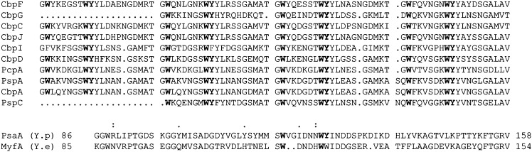
The pH 6 antigen (Psa) of Yersinia pestis consists of fimbriae that bind to two receptors: β1-linked galactosyl residues in glycosphingolipids and the phosphocholine group in phospholipids. Despite the ubiquitous presence of either moiety on the surface of many mammalian cells, Y. pestis appears to prefer interacting with certain types of human cells, such as macrophages and alveolar epithelial cells of the lung. The molecular mechanism of this apparent selectivity is not clear. Site-directed mutagenesis of the consensus choline-binding motif in the sequence of PsaA, the subunit of the Psa fimbrial homopolymer, identified residues that abolish galactosylceramide binding, phosphatidylcholine binding, or both. The crystal structure of PsaA in complex with both galactose and phosphocholine reveals separate receptor binding sites that share a common structural motif, thus suggesting a potential interaction between the two sites. Mutagenesis of this shared structural motif identified Tyr126, which is part of the choline-binding consensus sequence but is found in direct contact with the galactose in the structure of PsaA, important for both receptor binding. Thus, this structure depicts a fimbrial subunit that forms a polymeric adhesin with a unique arrangement of dual receptor binding sites. These findings move the field forward by providing insights into unique types of multiple receptor-ligand interactions and should steer research into the synthesis of dual receptor inhibitor molecules to slow down the rapid progression of plague.


Conflict of interest statement

The authors declare no conflict of interest.

Figures

Fig. 1.
Fig. 1.
Sequence motif of dscPsaA. Alignment of the amino acid sequences of PsaA of Y. pestis and MyfA of Y. enterocolitica with those of the choline-binding motifs of Streptococcus pneumoniae TIGR4 CBPs (21). Highly conserved residues of the motif are shown in bold. PCho and β1-linked galactose-binding residues of Psa are indicated by a period and a colon above the PsaA sequence, respectively.
Fig. 2.
Fig. 2.
Crystal structures of dscPsaA in complex with receptors. (A) A topological diagram of dscPsaA structure. (B) Ribbon representation of the structure of the ternary complex of dscPsaA with bound galactose and phosphocholine. All β-strands are shown in yellow except for the donor strand, which is shown in cyan, and labeled. All loops connecting pairs of β-strands are shown in green coils. Bound receptor molecules are shown as stick models with carbon atoms colored in magenta, nitrogen in blue, phosphorus in yellow, and oxygen in red. (C) Stereoscopic pair showing details of the galactose-binding niche. The bound galactose molecule and residues important for its binding are given as stick models and labeled. Potential hydrogen bonds are drawn as black lines. (D) Stereoscopic pair showing details of the phosphocholine-binding pocket. Phosphocholine and residues involved in its binding are shown as stick models.
Fig. 3.
Fig. 3.
Structural analysis of PsaA, its alignment with Cfa1 pilin, and functional implications. (A) Electrostatic potential surface of dscPsaA. The red and blue surfaces represent negative and positive potentials, respectively. White surface indicates a hydrophobic surface. Bound receptors are shown as stick models with the same color codes as in Fig. 2. (B) Structural alignment of dscPsaA and Caf1 pilin in the FGL family, showing as Cα tracing in green and yellow, respectively. (C) Model of Psa polyadhesin based on the structure of the F1 fimbriae (32). Seven subunits are shown, in which residues interacting with galactose are drawn as sphere models in light green and those interacting with PCho are colored in pink. Magnified portion shows the linkage between two neighboring subunits.
Fig. 4.
Fig. 4.
Changes in binding properties of WT and variant Psa. Binding of WT and substituted variants of Psa to PC (A) or GC (B) was evaluated by ELISA. The data are expressed as mean percent adhesion relative to the WT ± SE of three values and represent one of three reproducible experiments. *P < 0.05. Adhesion of E. coli SE 5000 expressing WT and substituted variants of Psa to A549 cells (C) or RAW 264.7 cells (D). Adhesion assays were carried out as described in Materials and Methods. Adherence percentages were calculated as the numbers of cell-associated bacteria divided by the total numbers of inoculated bacteria × 100 and were expressed as percent adhesion relative to the WT value (mean ± SE of three paired values that represent one of three reproducible experiments). All of the shown strains expressing mutated Psa bound significantly less well to both cell lines than the bacteria expressing WT Psa (P < 0.05).

Similar articles

Cited by

References

    1. Stenseth NC, et al. Plague: Past, present, and future. PLoS Med. 2008;5(1):e3. - PMC - PubMed
    1. Viboud GI, Bliska JB. Yersinia outer proteins: Role in modulation of host cell signaling responses and pathogenesis. Annu Rev Microbiol. 2005;59:69–89. - PubMed
    1. Felek S, Tsang TM, Krukonis ES. Three Yersinia pestis adhesins facilitate Yop delivery to eukaryotic cells and contribute to plague virulence. Infect Immun. 2010;78(10):4134–4150. - PMC - PubMed
    1. Zavialov A, Zav’yalova G, Korpela T, Zav’yalov V. FGL chaperone-assembled fimbrial polyadhesins: anti-immune armament of Gram-negative bacterial pathogens. FEMS Microbiol Rev. 2007;31(4):478–514. - PubMed
    1. Galván EM, Chen H, Schifferli DM. The Psa fimbriae of Yersinia pestis interact with phosphatidylcholine on alveolar epithelial cells and pulmonary surfactant. Infect Immun. 2007;75(3):1272–1279. - PMC - PubMed

Publication types

MeSH terms