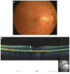
New optical coherence tomography fundus findings in a case of beta-thalassemia
- PMID: 23277736
- PMCID: PMC3532023
- DOI: 10.2147/OPTH.S35627
New optical coherence tomography fundus findings in a case of beta-thalassemia
Abstract
Patients with beta-thalassemia may present with an acquired diffuse elastic tissue defect due to degeneration of elastic tissue along with vaso-occlusive findings in the retinal microvasculature. Here we report the case of a patient with granular-like accumulation presenting as black sunburst lesions detected by optical coherence tomography (OCT). A 38-year-old man with beta-thalassemia intermedia associated with angioid streaks complained of deterioration of vision in both eyes. Funduscopic examination revealed small, round, hyperpigmented lesions bilaterally. During the early and late phases of fluorescein angiography, granular hyperfluoresence was present, associated with pigment decompensation and mottled-like hypofluorescence. The main OCT finding was the presence of granuloid-like accumulations at the retinal pigment epithelium level. Granule penetration was also noticed at the photoreceptor layer, while isolated granuloid-like accumulations were found in the inner layers of the macula and choroid. In this case, the new OCT finding was the granular-like hyperpigmented accumulations in the macula located at the level of the retinal pigment epithelium. To the best of our knowledge, our OCT findings show for the first time granuloid-like accumulations representing black sunburst lesions.
Keywords: angioid streaks; beta-thalassemia; granuloid-like accumulation; optical coherence tomography; retinal pigment epithelium.
Figures


Similar articles
-
Optical coherence tomographic features in a case of bilateral macular coloboma with strabismus.Eye Sci. 2011 Dec;26(4):244-6. doi: 10.3969/j.issn.1000-4432.2011.04.012. Eye Sci. 2011. PMID: 22187311
-
IMPROVED DETECTION AND DIAGNOSIS OF POLYPOIDAL CHOROIDAL VASCULOPATHY USING A COMBINATION OF OPTICAL COHERENCE TOMOGRAPHY AND OPTICAL COHERENCE TOMOGRAPHY ANGIOGRAPHY.Retina. 2019 Sep;39(9):1655-1663. doi: 10.1097/IAE.0000000000002228. Retina. 2019. PMID: 29927796
-
Contribution of OCT angiography in angioid streaks.J Fr Ophtalmol. 2021 Feb;44(2):209-217. doi: 10.1016/j.jfo.2020.04.056. Epub 2021 Jan 7. J Fr Ophtalmol. 2021. PMID: 33423815
-
Case report: the first case of unilateral retinal pigment epithelium dysgenesis in China.BMC Ophthalmol. 2020 Aug 20;20(1):340. doi: 10.1186/s12886-020-01609-4. BMC Ophthalmol. 2020. PMID: 32819322 Free PMC article.
-
[Pathophysiology of macular diseases--morphology and function].Nippon Ganka Gakkai Zasshi. 2011 Mar;115(3):238-74; discussion 275. Nippon Ganka Gakkai Zasshi. 2011. PMID: 21476310 Review. Japanese.
Cited by
-
Ocular abnormalities in beta thalassemia patients: prevalence, impact, and management strategies.Int Ophthalmol. 2020 Feb;40(2):511-527. doi: 10.1007/s10792-019-01189-3. Epub 2019 Oct 10. Int Ophthalmol. 2020. PMID: 31602527 Review.
-
Optical coherence tomography findings in patients with transfusion-dependent β-thalassemia.BMC Ophthalmol. 2022 Jun 24;22(1):279. doi: 10.1186/s12886-022-02490-z. BMC Ophthalmol. 2022. PMID: 35751049 Free PMC article.
-
Retinal abnormalities in β-thalassemia major.Surv Ophthalmol. 2016 Jan-Feb;61(1):33-50. doi: 10.1016/j.survophthal.2015.08.005. Epub 2015 Aug 29. Surv Ophthalmol. 2016. PMID: 26325202 Free PMC article. Review.
-
Spectral domain optical coherence tomography findings in Turkish sickle-cell disease and beta thalassemia major patients.J Curr Ophthalmol. 2019 Feb 14;31(3):275-280. doi: 10.1016/j.joco.2019.01.012. eCollection 2019 Sep. J Curr Ophthalmol. 2019. PMID: 31528761 Free PMC article.
-
Ophthalmic Evaluation in Beta-Thalassemia.Indian J Pediatr. 2017 Jul;84(7):509-514. doi: 10.1007/s12098-017-2339-8. Epub 2017 Apr 3. Indian J Pediatr. 2017. PMID: 28367614
References
-
- Aessopos A, Farmakis D, Loukopoulos D. Elastic tissue abnormalities resembling pseudoxanthoma elasticum in β thalassemia and the sickling syndromes. Blood. 2002;99:30–35. - PubMed
-
- Jensen OA. Bruch’s membrane in pseudoxanthoma elasticum. Histochemical, ultrastructural, and x-ray microanalytical study of the membrane and angioid streak areas. Graefes Arch Klin Exp Ophthalmol. 1977;203:311–320. - PubMed
-
- Aessopos A, Savvides P, Stamatelos G, et al. Pseudoxanthoma elasticum-like skin lesions and angioid streaks in beta-thalassemia. Am J Hematol. 1992;41:159–164. - PubMed
-
- Theodossiadis G, Ladas I, Koutsandrea C, Damanakis A, Petroutsos G. Thalassemia and macular subretinal neovascularization. J Fr Ophthalmol. 1984;7:115–118. French. - PubMed
-
- Aessopos A, Floudas SC, Kati M, et al. Loss of vision associated with angioid streaks in β thalassemia intermedia. Int J Hematol. 2008;87:35–38. - PubMed
Publication types
LinkOut - more resources
Full Text Sources

